Scientists Trigger Regeneration Pathway in Mammals Turning Scars Into Bone and Tendon
Biology

Scientists Trigger Regeneration Pathway in Mammals Turning Scars Into Bone and Tendon

Study finds mammals could reactivate dormant tissue regeneration when healing signals are precisely redirected, hinting at new regenerative therapies.

By Hassan Raza
Published:
Email this Article
Scientists Reveal Humans May Regrow Limbs Using Dormant Biological Code Scaled
Credit: Shutterstock | Dungrela Publishing

A recent report in Nature Communications shows that mammals can be coaxed into rebuilding complex structures such as bone, ligament, tendon and joint‑like tissue when wound‑healing signals are precisely timed. The work demonstrates that the default scar‑forming response can be overridden, hinting that regenerative pathways remain dormant rather than lost.

Why Mammals Scar Instead of Regrowing

For generations biologists have wondered why amphibians and fish can replace entire limbs while mammals, humans included, form scar tissue. Teams from Texas A &M University’s College of Veterinary Medicine and Biomedical Sciences approached the problem from a cellular‑flexibility perspective, suggesting that the key difference lies in the instructions given to repair cells rather than in an absent capability.

“Why some animals can regenerate and others, particularly humans, can’t is a big question that has been asked since Aristotle,” said Dr. Ken Muneoka, a professor in the VMBS’ Department of Veterinary Physiology & Pharmacology (VTPP). “I’ve spent my career trying to understand that.”

Muneoka and colleagues frame the issue as a biological switch that may still operate in mammals, albeit in a suppressed state. Unlocking that switch could transform strategies for wound repair and tissue engineering.

41467 2026 72066 Fig1 Html
A–C Representative longitudinal sections and μCT images (insets) of control and FGF2 treated digits 21 days post treatment (DPT). A Control BSA treated digits do not display a regenerative response and are truncated. B The majority of FGF2 treated digits are truncated without displaying a regenerative response. C A minority of FGF2 treated digits produce an ectopic skeletal element distal to the amputation that articulates with the stump bone. D Length measurements of the P2 stump 21 DPT (n = 52) indicated that FGF2 does not stimulate skeletal elongation compared to BSA treated controls (n = 22; unpaired t-test, ns > 0.05). E–G Representative longitudinal sections identifying EdU incorporation associated with bead implantation (*) at 4 DPT. E Few EdU positive cells are labeled following BSA treatment (n = 8). F EdU labeled cells are moderately enhanced following BMP2 treatment (n = 8). G EdU labeled cells are abundant following FGF2 treatment (n = 8). H Quantification of EdU labeled cells associated with the implanted bead following BSA, BMP2 and FGF2 treatment (n = 8 for each treatment; unpaired t-test, **p < 0.01, *p < 0.05). I qRT-PCR analysis of blastema-related transcripts and chondrogenic genes at 3 and 5 DPT following FGF2 treatment indicates that blastema-related genes are up‑regulated while chondrogenic genes are initially down‑regulated when compared to control BSA treated digits. FGF2: n = 16 digits pooled/time point (4 mice); BSA: n = 16 digits pooled/time point (4 mice). Unpaired t-test; ****p < 0.0001, ***p < 0.001, **p < 0.01, *p < 0.05. Error bars indicate standard deviation. J, K Immunostaining studies indicate a high level of Cxcr4 positive cells (n = 3) associated with the FGF2 bead (J) compared to controls (n = 3) (K). L, M In situ hybridization studies indicate an up‑regulation of Msx1 transcripts (n = 6) associated with blastema formation in the dorsal region of the stump (L) compared to controls (n = 4) (M). Credit: Nature Communications

Fibroblasts: Cells with Two Possible Paths

Fibroblasts normally rush to seal wounds, laying down collagen that becomes scar tissue. The new experiments reveal that these same cells can be redirected toward a blastema‑like state—a regenerative precursor—when exposed to a defined sequence of growth factors.

“It’s as if these cells can move in two different directions,” Muneoka said. “They could either make a scar or make a blastema. Our research focused on redirecting the behavior of fibroblasts already present at the injury site.”

Timing proved crucial: delivering the first signal too early or too late altered the outcome, indicating a narrow biological window during which cells remain receptive to a regenerative cue.

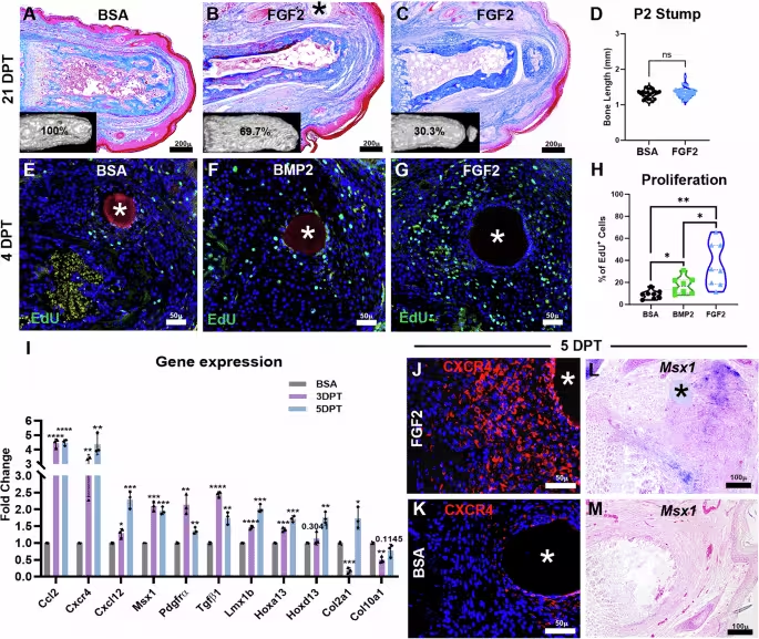
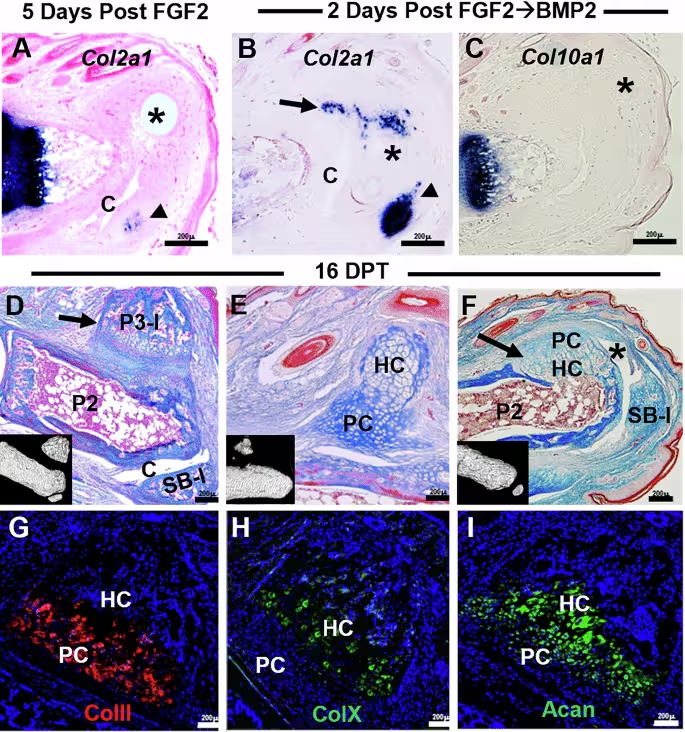
Guided Growth Factors Trigger Partial Regrowth

The protocol began with a factor that encouraged blastema formation, followed by a second cue that steered the nascent tissue toward organized bone, tendon, ligament and joint elements. Although the regenerated structures were not perfect copies of the original anatomy, they displayed functional architecture not typically seen in mammalian repair.

“We regenerated what you would expect to see at that level of injury,” Muneoka said. “The structures are there — just not in a perfect form.”

41467 2026 72066 Fig3 Html
A Unamputated digit showing the P2, P3, and sesamoid bone (SB). B In vivo imaging of the same digit 16 and 45 days post FGF2→BMP2 treatment (DPT) showing the regeneration of a single dorsal skeletal element that appears similar to a P3 bone (P3-l). C In vivo imaging of the same digit at 16 DPT and 45 DPT showing the regeneration of a dorsal P3-like skeletal element (P3-l) and a distal‑ventral element (SB-l) that is similar to a sesamoid bone. D P2 bone volume at 45 DPT indicate a significant increase of FGF2→BMP2 treated digits compared to BSA controls but does not reach the bone volume of unamputated digits (1‑way ANOVA with Tukey’s multiple comparison test; ****p < 0.0001). E P2 bone length indicate a significant increase in bone length induced by FGF2→BMP2 treatment compared to BSA controls but does not reach the bone length of unamputated digits (1‑way ANOVA with Tukey’s multiple comparison test; ****p < 0.0001). F All ectopic skeletal elements were spatially plotted onto an amputated P2 stump showing clustering at dorsal and distal/ventral positions at 16 DPT and 45 DPT. G Bone volume measurements of ectopic elements showing size differences between dorsal and distal‑ventral elements at 16 DPT and 45 DPT (2‑way ANOVA with Fisher’s LSD; ****p < 0.0001, *p < 0.05). H Histological section of a representative digit (inset) at 45 DPT that regenerated a dorsal P3‑l bone and a distal/ventral SB‑l element (n = 5). The P3‑l bone has a vascularized bone marrow cavity that is distinct from the SB‑l element that lacks a marrow cavity. Regenerated ligaments (L) and tendon (T) make attachments to the ectopic bones and a tendon‑bone enthesis (En) is circled. Regions of articular cartilage (AC) are identified based on histological staining (arrow). I Morphometric PCA results show that regenerated dorsal ectopic elements (solid gray) cluster with unamputated P3 bones (open gray), while regenerated distal‑ventral elements (solid black) cluster with unamputated sesamoid bones (open black). Gray lines denote K‑means decision boundaries (k = 4), whereas the red dashed line denotes the decision boundary for k = 2. Statistics: *P < 0.05; **P < 0.01; ***P < 0.001; ****P < 0.0001.Credit: Nature Communications

Clinical Outlook and Next Steps

The authors caution that full limb regeneration remains out of reach for humans, but the ability to steer scar formation toward more functional tissue could reshape postoperative care and chronic wound management. The study’s senior authors suggest that applying these molecular cues during the normal healing process may already confer measurable benefits.

“The cells that we thought to be unprogrammable, in fact are,” Suva said. “The capacity is not absent — it’s just obscured.” He also noted, “People should start thinking about using these signals during the healing process,” Muneoka said. “Even shifting the response slightly away from scarring could have real benefits.”

“This changes the way we think about what’s possible,” Suva added. “Once you show that regeneration can be activated, it opens the door to asking entirely new questions.” The findings, published in Nature Communications, present a model for probing latent regenerative capacity in mammals and lay groundwork for future therapeutic exploration.

Fact Checked

This article has been fact checked for accuracy, with information verified against reputable sources. Learn more about us and our editorial process.

Last reviewed on .

Article history

  • Latest version

Reference(s)

  1. Yu, Ling. “Digit regeneration in mice is stimulated by sequential treatment with FGF2 and BMP2 - Nature Communications.”, vol. 17, no. 1, April 17, 2026, pp. 5346 Nature, doi: 10.1038/s41467-026-72066-8. <https://www.nature.com/articles/s41467-026-72066-8>.

Cite this page:

Raza, Hassan. “Scientists Trigger Regeneration Pathway in Mammals Turning Scars Into Bone and Tendon.” BioScience. BioScience ISSN 2521-5760, 19 June 2026. <https://www.bioscience.com.pk/en/subject/biology/scientists-reveal-humans-may-regrow-limbs-using-dormant-biological-code>. Raza, H. (2026, June 19). “Scientists Trigger Regeneration Pathway in Mammals Turning Scars Into Bone and Tendon.” BioScience. ISSN 2521-5760. Retrieved June 19, 2026 from https://www.bioscience.com.pk/en/subject/biology/scientists-reveal-humans-may-regrow-limbs-using-dormant-biological-code Raza, Hassan. “Scientists Trigger Regeneration Pathway in Mammals Turning Scars Into Bone and Tendon.” BioScience. ISSN 2521-5760. https://www.bioscience.com.pk/en/subject/biology/scientists-reveal-humans-may-regrow-limbs-using-dormant-biological-code (accessed June 19, 2026).

Follow us on social media

End of the article