Lab‑Grown Human Embryo Models Reach Gastrulation and Spark Early Organ Formation
Researchers have mimicked a key early‑human development stage, paving the way for lab‑grown, transplantable organs in a breakthrough study.
Researchers in China have reported a major advance in developmental biology: laboratory‑grown human embryo analogues that progress through gastrulation and generate the first cell types that will form major organs. The study, appearing in Cell, provides a powerful tool for probing a developmental stage that has been largely inaccessible to scientists.
Breakthrough in Modeling Early Human Development
Gastrulation, a fleeting but pivotal phase in which a simple cell layer folds into a three‑dimensional structure that sets the blueprint for the entire body, has long eluded direct observation because international guidelines forbid culturing human embryos past 14 days after fertilization. The new work overcomes this limitation by engineering embryo‑like constructs that mimic the natural process.
“Gastrulation is when the body’s basic architecture is established, transforming the embryo from a flat disc into a three-dimensional structure,” says Yu Leqian, corresponding author and professor at the Institute of Zoology, Chinese Academy of Sciences.
Earlier in‑vitro models captured only fragments of early development and failed to form a primitive streak—a structure that orchestrates cell movement, specialization, and organ precursor formation. By incorporating a primitive‑streak‑like element, the new models achieve a level of organization previously unattainable.
Spatial‑Biology Approach Drives Organized Development
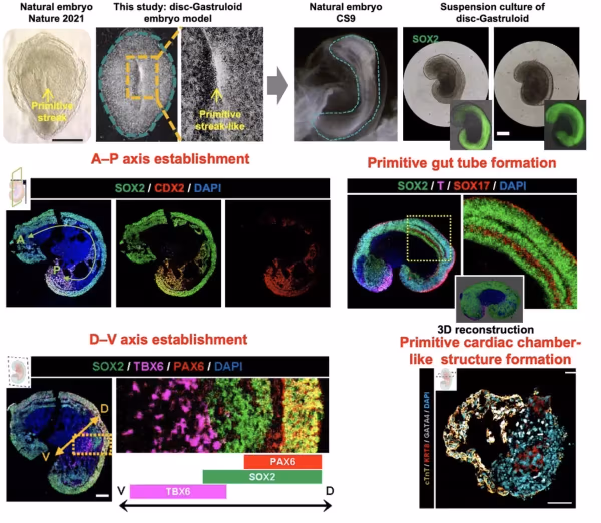
Instead of relying on the spontaneous self‑assembly of stem cells, the team applied spatial‑biology techniques to arrange early human cells in defined patterns. The resulting “disc‑Gastruloids” displayed gastrulation‑like behavior in more than 80 % of cases, forming structures that resembled a primitive streak.
Following this milestone, the constructs showed coordinated cell migration across their surface, mirroring events seen in authentic embryos. Subsequent stages included formation of neural tube‑like tissue, primitive gut, early lung, liver and pancreas progenitors, and a beating cardiac chamber. Single‑cell profiling revealed that the cellular composition of the models closely matched that of human embryos at roughly 21 days, representing one of the most accurate in‑vitro recapitulations to date.
Implications for Regenerative Medicine and Disease Modeling
While the prospect of transplantable lab‑grown organs remains distant, reproducing gastrulation tackles a core challenge: delivering not just the right cell types but also the developmental cues that guide their assembly into functional tissues. The ability to generate early “organ‑seed” cells in a setting that mimics natural development could eventually enhance the precision of tissue engineering efforts.
“The study has laid the foundation for the ultimate goal of large-scale, modular production of organ‑seed cells in vitro to support organ manufacturing and regenerative medicine – potentially enabling tissue repair or even the construction of organs in the laboratory,” Yu explains.
Beyond organ fabrication, the models offer a versatile system for investigating congenital anomalies, early‑onset developmental disorders, and the molecular pathways that drive organogenesis, potentially informing new therapeutic strategies and reducing dependence on donor organs.
This article has been fact checked for accuracy, with information verified against reputable sources. Learn more about us and our editorial process.
Last reviewed on .
Article history
- Latest version
Cite this page:
- Posted by Hassan Raza