Kissing Bug Cell Lines Reveal Hidden Genetics Behind Chagas Disease Vector
Comprehensive RNA sequencing of stable embryo cultures uncovers active signaling pathways and offers a new experimental platform for vector biology.
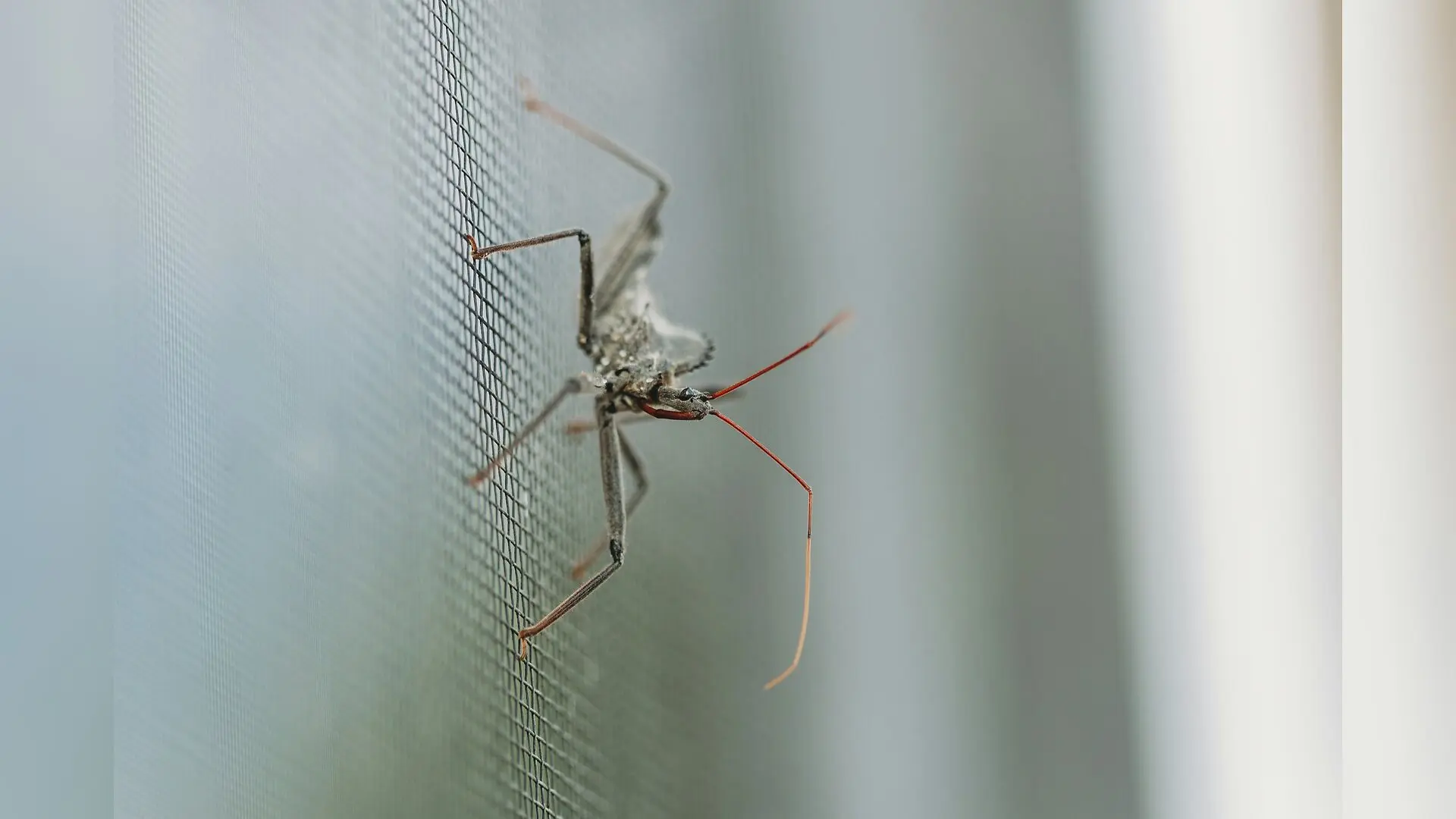
Two stable cell lines derived from kissing bug embryos reveal surprisingly active genetic machinery, offering new ways to study the parasite carrier responsible for Chagas disease. These insect cell lines, named RPE/LULS53 and RPE/LULS57, represent the first stable cultures obtained from a blood-sucking insect of the hemimetabolous group, which undergoes incomplete metamorphosis.
Researchers mapped the active genes inside these cultures, providing a clearer look at the molecular inner workings of a major disease vector. The detailed findings were published in PLOS Neglected Tropical Diseases in November 2026 by Laura de Andrade Tavares and colleagues.
What Transformed Gene Sequencing Reveals About Kissing Bug Cells
To uncover the molecular profiles of these cellular tools, the research team extracted ribonucleic acid, which carries genetic instructions from DNA, and sequenced it using advanced high-throughput laboratory platforms. Around 26 million paired-end reads were generated for each cell line to measure gene activity accurately. When aligned to the reference genome of Rhodnius prolixus, over 85 percent of the genetic sequences matched successfully, confirming that the cells retain their authentic insect identity.
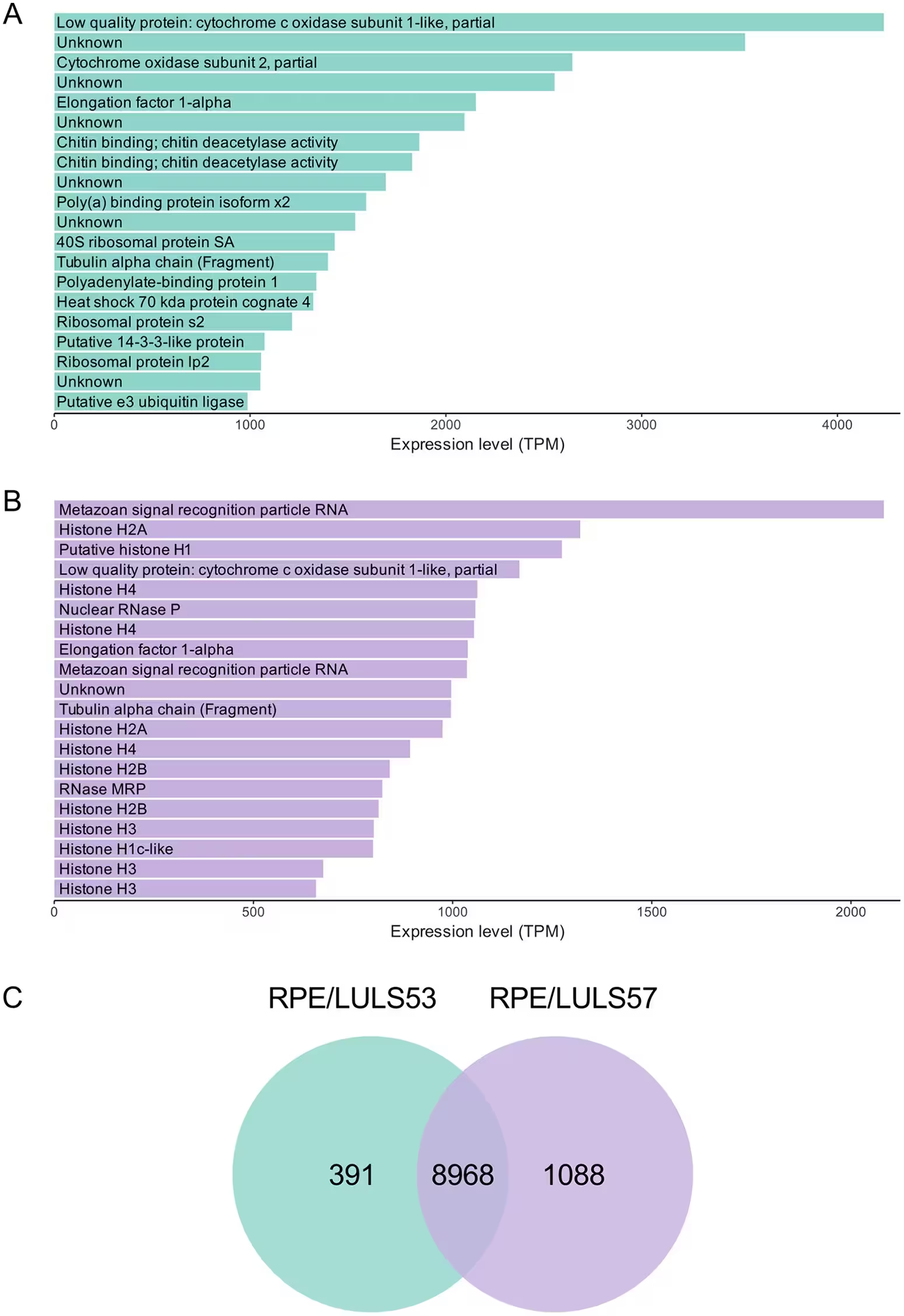
The analysis showed that over 8,900 genes are actively expressed in both cell lines, sharing a common core of metabolic and structural functions. Highly abundant transcripts included genes responsible for protein synthesis, energy production via cytochrome c oxidase, and cellular scaffolding through tubulin and elongation factors. Despite these shared traits, the two lines showed distinct expression patterns that point toward functional divergence.
Which Pathways and Defense Mechanisms Stand Out in Each Line
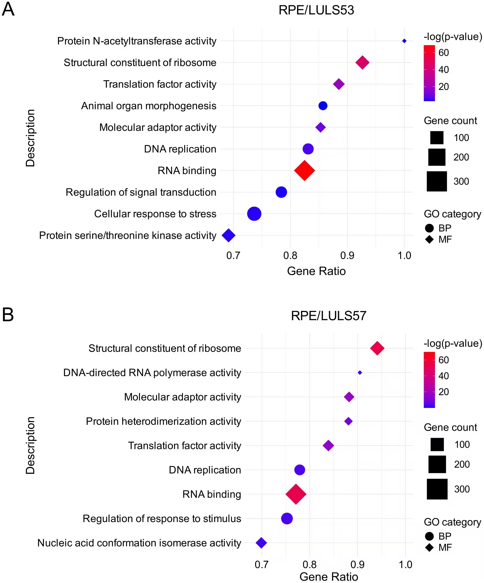
Gene Ontology enrichment tests highlighted different functional priorities between the two cultures. The RPE/LULS53 cell line showed higher activity in genes linked to signal transduction, stress responses, and animal organ morphogenesis. In contrast, RPE/LULS57 concentrated more heavily on transcription, translation, and nucleic acid metabolism, alongside significant expression of histone-encoding genes and specialized small non-coding ribonucleic acids.
Because these insects feed exclusively on blood, researchers also examined the management of reactive oxygen species and iron handling. Genes associated with redox homeostasis, including dual oxidases and catalases, were more abundant in RPE/LULS53. Furthermore, the team detected transcripts from numerous transposable elements, which are segments of DNA that change their position within the genome. These included both ancient resident transposons and horizontally transmitted elements acquired from other animal species over millions of years.
How This Genetic Map Enables Future Laboratory Experiments
To demonstrate practical applications for these cell lines, the investigators tested whether natural promoter sequences could drive foreign gene expression in culture. They successfully isolated a roughly 500-base-pair regulatory region from the beta-tubulin gene, which displays high natural expression in both cell lines. By attaching this promoter upstream of a green fluorescent protein reporter sequence, the team achieved clear fluorescence in RPE/LULS57 cells following transient transfection.
This proof of concept confirms that the transcriptomic data can guide the design of functional expression systems for genetic manipulation. Because classical genetic tools such as stable mutant lines remain difficult to establish in whole kissing bugs due to their lengthy six-month life cycle, these validated cell lines provide a much-needed platform. They will help scientists dissect cellular signaling, antiviral immunity, and the complex biological interactions between the insect vector, endogenous viruses, and the protozoan parasite Trypanosoma cruzi.
The research was published in PLOS Neglected Tropical Diseases on November 16, 2026.
This article has been fact checked for accuracy, with information verified against reputable sources. Learn more about us and our editorial process.
Last reviewed on .
Article history
- Latest version
Reference(s)
- de Andrade Tavares, Laura., et al. “Transcriptomic profiling of embryo-derived cell lines from the Chagas disease insect vector Rhodnius prolixus (Hemiptera: Triatominae).” PLOS Neglected Tropical Diseases, vol. 20, no. 11, November 16, 2026, pp. e0014346 Public Library of Science (PLoS), doi: 10.1371/journal.pntd.0014346. <https://doi.org/10.1371/journal.pntd.0014346>.
Cite this page:
- Posted by Elizabeth Taylor