Massive 400-Kilodalton Enzyme Structure Reveals Hidden Off Switch
Genetics

Massive 400-Kilodalton Enzyme Structure Reveals Hidden Off Switch

High-resolution imaging shows how tiny structural shifts far from active sites can control the breakdown of essential amino acids.
By Faisal Qureshi
Published:
Email this Article
Massive 400-Kilodalton Enzyme Structure Reveals Hidden Off Switch
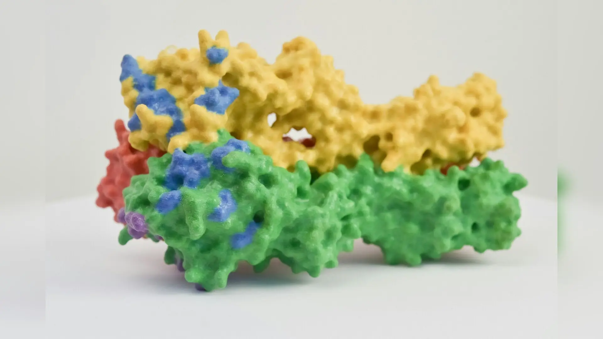
Photo by National Institute of Allergy and Infectious Diseases on Unsplash

Tiny structural shifts on a massive 400-kilodalton enzyme can completely alter how human cells break down an essential amino acid, according to a structural biology study published in Science Advances.

The research team mapped the full-length architecture of 2-aminoadipic semialdehyde synthase, a large protein complex known as AASS that controls the initial, rate-limiting steps of lysine degradation. By capturing high-resolution images of the enzyme bound to various natural substrates and products, the investigators revealed a surprisingly dynamic architecture. The complex features a central tetrameric core surrounded by flexible catalytic domains that move freely to pass chemical intermediates between active sites without letting them escape.

How Remote Residues Control Enzyme Activity

The scientists focused on an unexpected regulatory site located more than 12 angstroms away from the main active pocket. Through biochemical assays and molecular dynamics simulations, they discovered that modifying a single amino acid at position 414, specifically a cysteine residue, triggers widespread conformational changes across the protein. Even though this site sits far from the active center, altering it shifts the enzyme’s optimal pH and dramatically enhances its affinity for essential cosubstrates like NADPH.
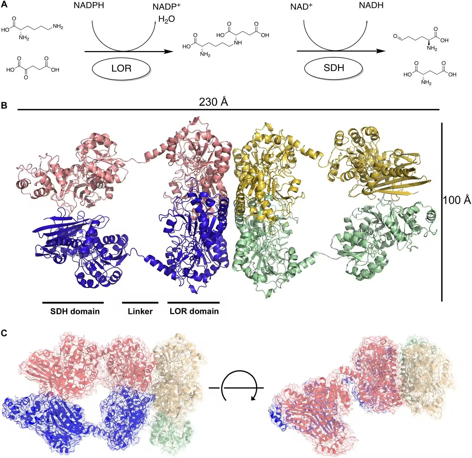
Fig. 1: Overall structure and function of AASS. (A) Enzyme reaction of LOR and SDH domains of AASS. (B) Overall structure of AASS tetrameric complex. The complex is colored by chain, with the two domains indicated along with the helical linker connecting them. (C) High-resolution structure of the tetrameric complex, with six of the eight domains visible. The electron density is shown colored by the corresponding chains and rotated to show two different perspectives. The transparent map is overlaid with the model.
Overall structure and function of AASS. (A) Enzyme reaction of LOR and SDH domains of AASS. (B) Overall structure of AASS tetrameric complex. The complex is colored by chain, with the two domains indicated along with the helical linker connecting them. (C) High-resolution structure of the tetrameric complex, with six of the eight domains visible. The electron density is shown colored by the corresponding chains and rotated to show two different perspectives. The transparent map is overlaid with the model. Credit: Ruoxi Wu et al. / Science advances

Further investigation showed that these distant modifications disrupt a crucial electrostatic network inside the protein. In the native enzyme, a stabilizing interaction locks two mobile lobes together. When the cysteine at position 414 undergoes chemical modification or mutation, that internal latch loosens, granting the protein structural flexibility that favors a more active catalytic state.

Potential Implications for Rare Metabolic Disorders

Understanding how to turn this metabolic pathway on and off carries significant clinical relevance. Defects in enzymes that function immediately downstream of AASS lead to severe neuro-metabolic conditions like glutaric aciduria type 1 and pyridoxine-dependent epilepsy, both of which lack Food and Drug Administration-approved treatments. Because harmless genetic deficiencies in AASS itself suggest that safely inhibiting the enzyme is possible, researchers have pursued it as a therapeutic strategy to prevent the buildup of toxic metabolites.
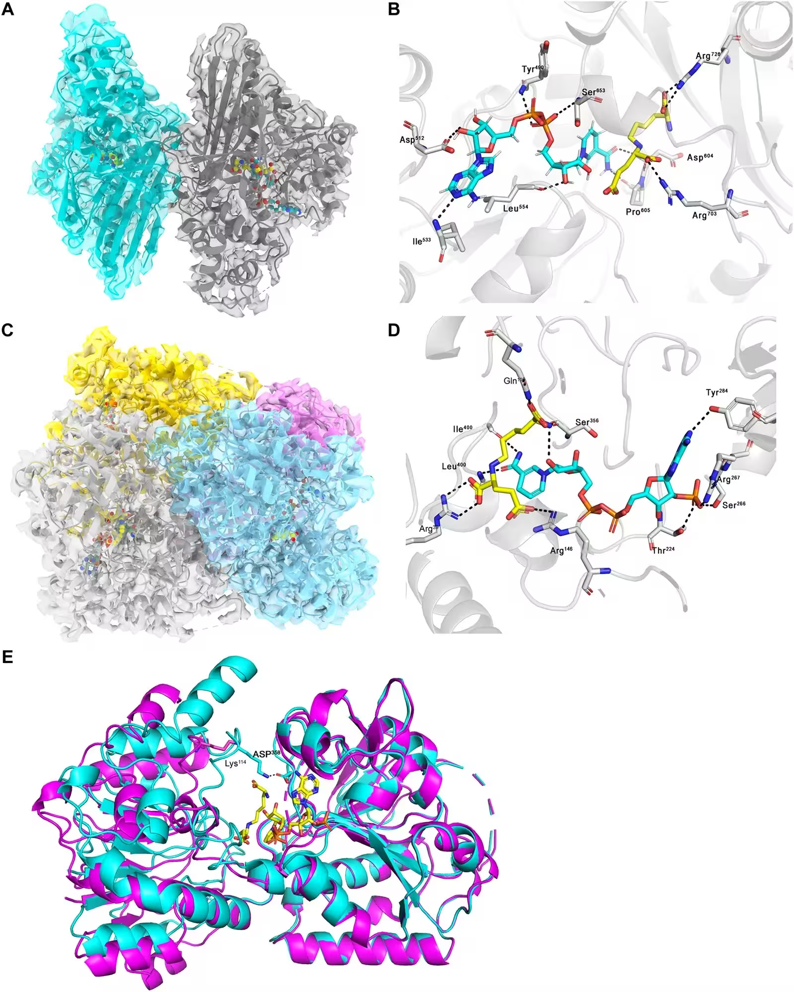
Fig. 2: Structure of the individual domains of AASS. (A) Overall structure of the SDH domain, shown as the dimer and colored by chain, overlaid on the cryo-EM map. NADH and saccharopine are shown as spheres in the active site. (B) Close-up of SDH active site. NADH is shown in blue sticks, saccharopine in yellow. Putative contacts are shown with dashed lines to indicated residues. (C) Overall structure of the LOR domain, shown as the tetramer and colored by chain, overlaid on the cryo-EM map. NADPH and saccharopine are shown as spheres in the active site. (D) Close-up of LOR active site. NADPH is shown in blue sticks, saccharopine in yellow. Putative contacts are shown with dashed lines to indicated residues. (E) Overlay of apo crystal structure (PDB 8E8U, magenta) with the new cryo-EM complex (blue), with substrates (yellow) and key residues (cyan) shown as sticks.
Structure of the individual domains of AASS. (A) Overall structure of the SDH domain, shown as the dimer and colored by chain, overlaid on the cryo-EM map. NADH and saccharopine are shown as spheres in the active site. (B) Close-up of SDH active site. NADH is shown in blue sticks, saccharopine in yellow. Putative contacts are shown with dashed lines to indicated residues. (C) Overall structure of the LOR domain, shown as the tetramer and colored by chain, overlaid on the cryo-EM map. NADPH and saccharopine are shown as spheres in the active site. (D) Close-up of LOR active site. NADPH is shown in blue sticks, saccharopine in yellow. Putative contacts are shown with dashed lines to indicated residues. (E) Overlay of apo crystal structure (PDB 8E8U, magenta) with the new cryo-EM complex (blue), with substrates (yellow) and key residues (cyan) shown as sticks. Credit: Ruoxi Wu et al. / Science advances

To explore natural regulatory molecules, the team screened a library containing 980 endogenous metabolites and identified several compounds capable of modulating AASS activity. Among them, 1-methylnicotinamide, a byproduct of vitamin B3 breakdown, acted as an inhibitor by triggering the formation of a selective disulfide bridge between two active-site cysteines. This reversible locking mechanism provides cells with a way to tune lysine catabolism based on metabolic demands and cellular stress.

While the study demonstrates how subtle chemical tweaks can dramatically influence complex protein machinery, the authors note that further work is required to determine whether these endogenous metabolites regulate the pathway in living organisms under physiological conditions.

The research was published in Science advances on November 13, 2026.

Fact Checked

This article has been fact checked for accuracy, with information verified against reputable sources. Learn more about us and our editorial process.

Last reviewed on .

Article history

  • Latest version

Reference(s)

  1. Wu, Ruoxi., et al. “Structural and biochemical insight into allosteric regulation of the human 2-aminoadipic semialdehyde synthase.” Science Advances, vol. 12, no. 46, November 13, 2026 American Association for the Advancement of Science (AAAS), doi: 10.1126/sciadv.aej2268. <https://doi.org/10.1126/sciadv.aej2268>.

Cite this page:

Qureshi, Faisal. “Massive 400-Kilodalton Enzyme Structure Reveals Hidden Off Switch.” BioScience. BioScience ISSN 2521-5760, 16 November 2026. <https://www.bioscience.com.pk/en/subject/genetics/structural-and-biochemical-insight-into-allosteric-regulation-of-the-human-2-aminoadipic-semialdehyde-synthase>. Qureshi, F. (2026, November 16). “Massive 400-Kilodalton Enzyme Structure Reveals Hidden Off Switch.” BioScience. ISSN 2521-5760. Retrieved November 16, 2026 from https://www.bioscience.com.pk/en/subject/genetics/structural-and-biochemical-insight-into-allosteric-regulation-of-the-human-2-aminoadipic-semialdehyde-synthase Qureshi, Faisal. “Massive 400-Kilodalton Enzyme Structure Reveals Hidden Off Switch.” BioScience. ISSN 2521-5760. https://www.bioscience.com.pk/en/subject/genetics/structural-and-biochemical-insight-into-allosteric-regulation-of-the-human-2-aminoadipic-semialdehyde-synthase (accessed November 16, 2026).
End of the article