Human Chromosome Length Scales With DNA Content in Strikingly Linear Fashion
A new mathematical unit bridges the gap between microscopic chromosome lengths and molecular DNA folding models.

Human metaphase chromosome length scales almost perfectly with DNA content, following a remarkably steady mathematical rule across all 24 human chromosomes.
Researchers Michell Goyal and Ravi Goyal pooled cytogenetic measurements from five historical studies of cultured peripheral lymphocytes and compared them against finished human reference genome assemblies. Their analysis, published in PLOS One, reveals a linear scaling relationship that bridges the gap between microscopic chromosome lengths and molecular Hi-C structural models of DNA folding.
How Much DNA Fits Into a Micron of Chromosome Length?
When cells prepare to divide during mitosis, their sprawling genetic material condenses into distinct, visible structures known as chromosomes. By analyzing average chromosome lengths ranging from 1.58 micrometers for chromosome 21 up to 8.21 micrometers for chromosome 1, the authors found that length increases linearly with base-pair count at a compaction density of 33.4 nanometers per megabase.
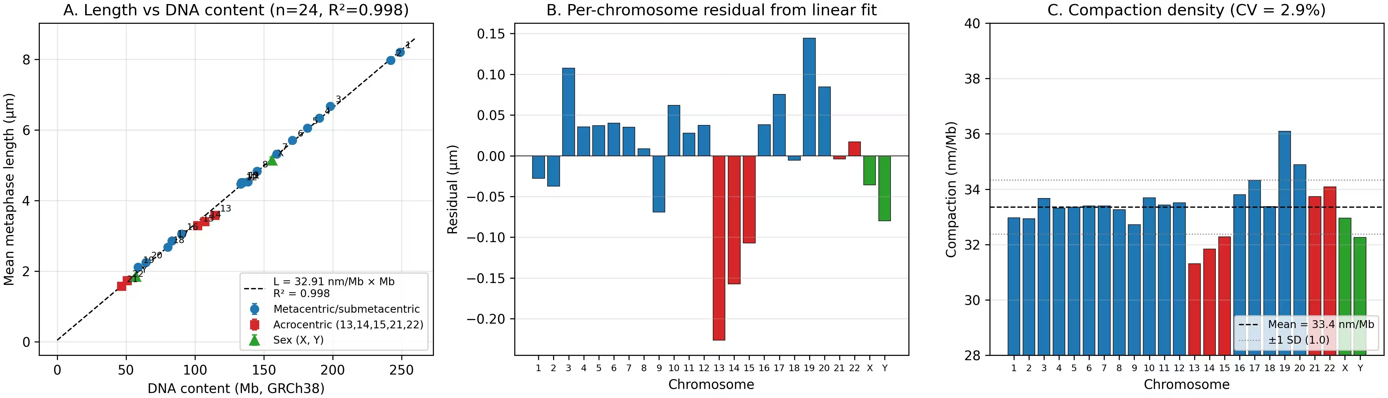
This tight correlation yields an R-squared value of 0.998, indicating that variation in DNA content explains nearly all differences in chromosome length. The team tested whether chromosomes are built from discrete folding blocks, but found no statistical support for a specific quantum size. Instead, the data point to a smooth, proportional compaction process across the entire human genome.
What Is the Metaphase Chromatin Unit and Why Does It Matter?
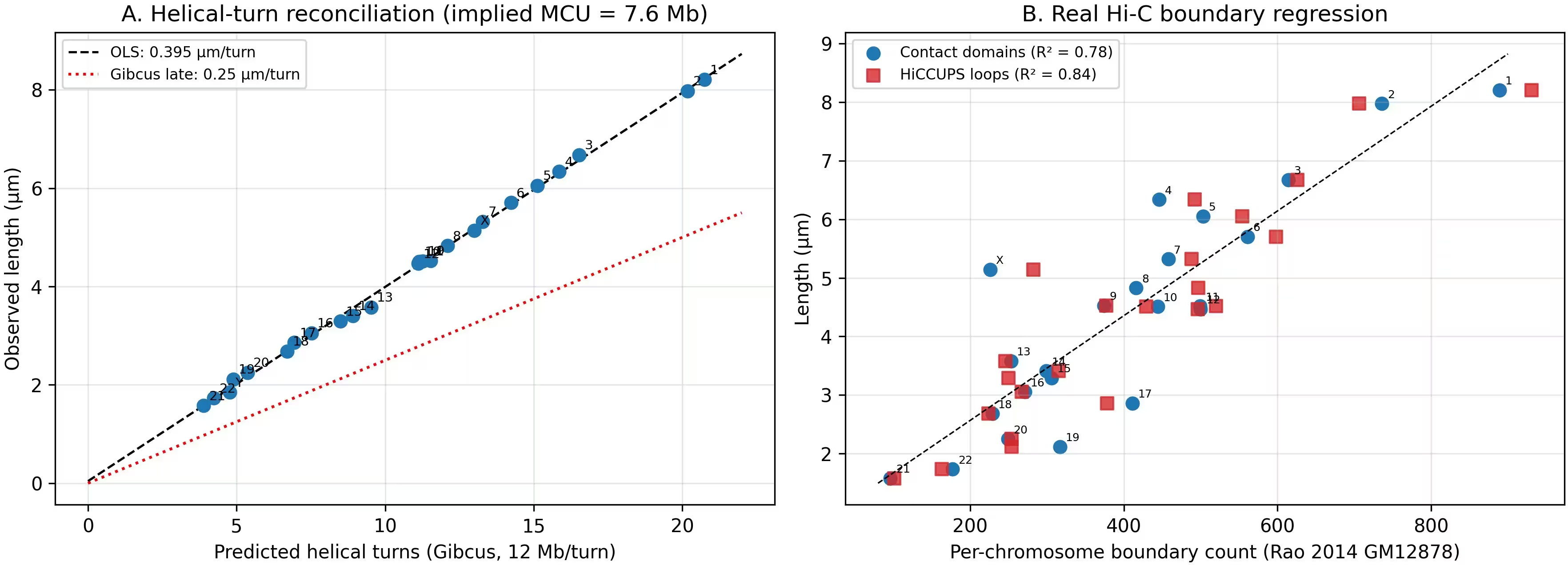
To connect these macro-scale cytogenetic measurements with modern biophysics, the researchers proposed an operational benchmark called the Metaphase Chromatin Unit, or MCU. Under this definition, 1 MCU corresponds to 7.6 megabases of DNA compacted into 0.25 micrometers of axial length.
This specific dimension matches one late-metaphase helical turn described in previous high-resolution folding models. Across the entire human haploid genome, this framework accounts for roughly 406 MCUs, matching the total observed mitotic length of approximately 102.7 micrometers. Individual chromosomes span from 6.1 MCUs in the smallest autosome up to 32.8 MCUs in chromosome 1.
Can This Geometric Scaling Predict Clinical Genetic Disorders?
The 33.4 nanometers per megabase compaction rate also sheds light on clinical diagnostics. By factoring in the Abbe optical diffraction limit of standard light microscopy, which sits near 196 nanometers, the researchers calculated a cytogenetic detection threshold of approximately 6 megabases.
When applied to well-characterized microdeletion syndromes, this optical threshold successfully classifies nine out of nine clinical conditions as either requiring specialized molecular testing like fluorescence in situ hybridization or remaining visible under standard karyotype analysis. Disorders involving smaller segments, such as Williams syndrome at 1.6 megabases and DiGeorge syndrome at 3 megabases, fall below the optical limit, whereas larger deletions are reliably detected by eye.
What Limitations Affect These Chromosome Models?
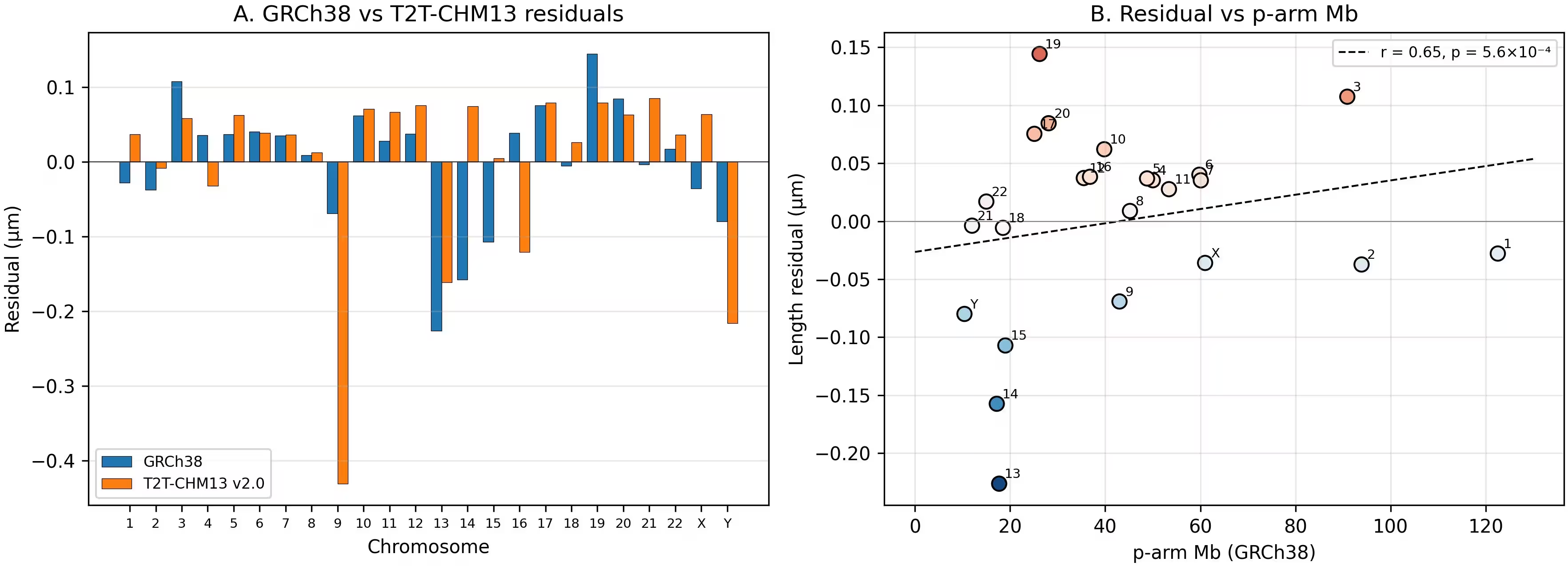
The authors note several caveats regarding their pooled dataset. The five cytogenetic studies spanned several decades, from 1964 to 2013, involving varying fixation protocols and potential inter-laboratory variations. Furthermore, reanalysis using the T2T-CHM13 genome assembly revealed that heavy concentrations of pericentromeric satellite DNA undergo extreme over-condensation, showing compaction densities at least eight times higher than the chromosome-wide average.
Despite these local variations, the overarching linear model provides a quantitative anchor. By tying together microscopic chromosome dimensions, Hi-C boundary counts, and clinical deletion limits, the work offers a unified mathematical reference for human chromosomal architecture.
The research was published in PLOS One on November 13, 2026.
This article has been fact checked for accuracy, with information verified against reputable sources. Learn more about us and our editorial process.
Last reviewed on .
Article history
- Latest version
Reference(s)
- Goyal, Michell., et al. “The metaphase chromatin unit: A novel unit of higher-order chromosome organization in human mitotic cells.” PLOS One, vol. 21, no. 11, November 13, 2026, pp. e0361508 Public Library of Science (PLoS), doi: 10.1371/journal.pone.0361508. <https://doi.org/10.1371/journal.pone.0361508>.
Cite this page:
- Posted by Faisal Qureshi