Immune Cell Growth Factors Dictate Chikungunya Virus Susceptibility
Laboratory experiments reveal why certain macrophages support viral replication while others mount a robust defense
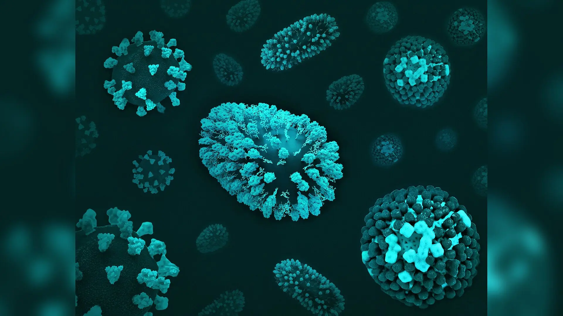
Human immune cells grown in the laboratory show wildly different susceptibility to Chikungunya virus depending on the chemical signals that shaped their early development. The mosquito-borne pathogen, which causes persistent joint pain in infected individuals, successfully replicates in immune cells primed by one major growth factor but fails to multiply in cells guided by another.
The research team, led by scientists at the Icahn School of Medicine at Mount Sinai, investigated how monocyte-derived macrophages respond to infection when differentiated using distinct colony-stimulating factors. Monocytes are white blood cells that circulate in the bloodstream before migrating into tissues, where they mature into macrophages. This maturation process can be nudged in different directions by local signaling proteins.
How Growth Factors Change Viral Susceptibility
When the researchers exposed human macrophages grown with granulocyte-macrophage colony-stimulating factor to Chikungunya virus, the cells supported active viral replication and expressed high levels of viral proteins. In contrast, cells differentiated using macrophage colony-stimulating factor resisted viral replication and showed no detectable viral proteins.
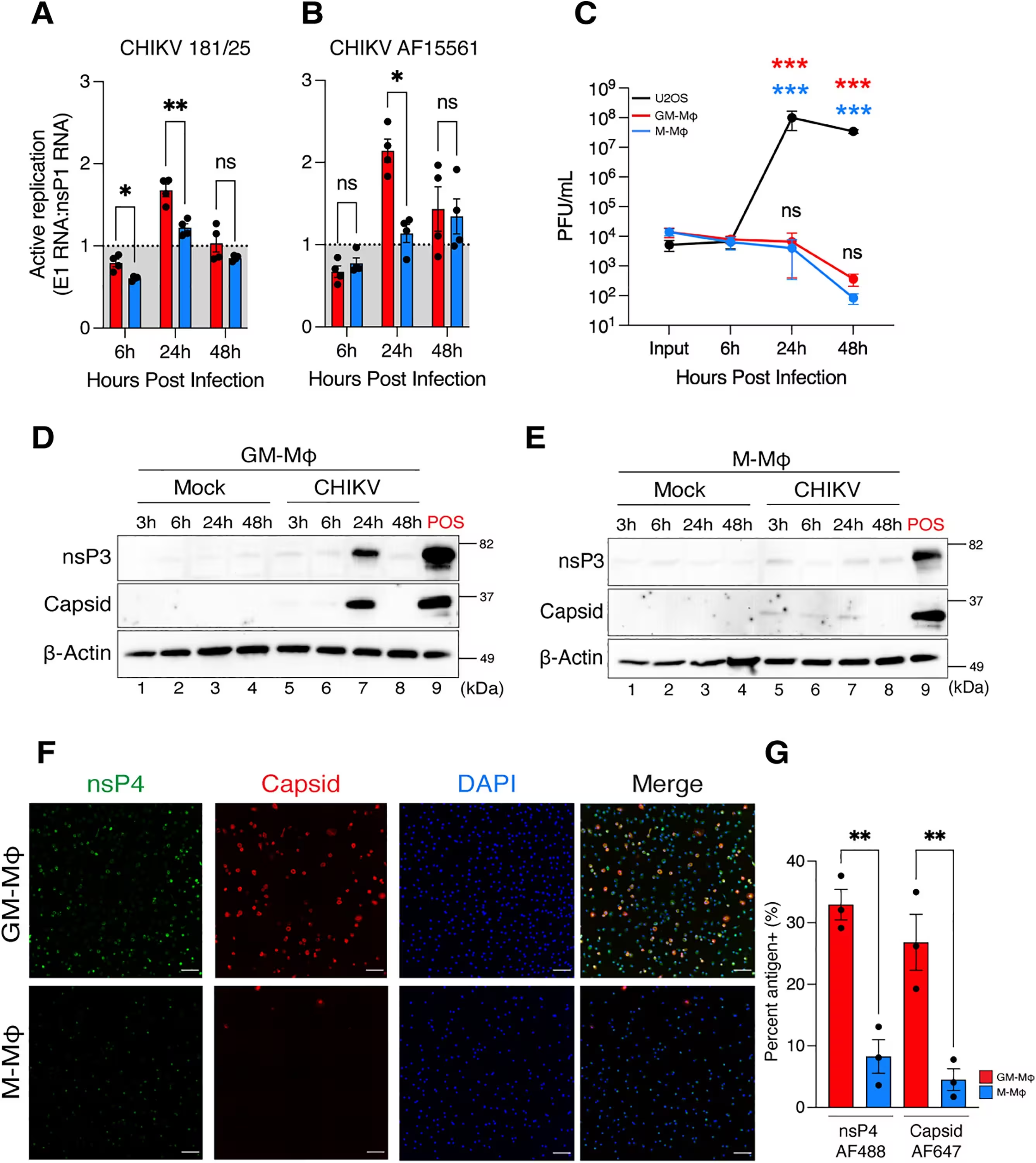
The study, published in PLOS Pathogens, points out that despite these divergent replication outcomes, both types of macrophages adopted a pro-inflammatory activation state after exposure. The researchers also tested Mayaro virus, a related arthritogenic alphavirus that shares receptors with Chikungunya, and observed a similar preference for infecting cells primed by granulocyte-macrophage colony-stimulating factor.
Behind the Post-Entry Cellular Resistance
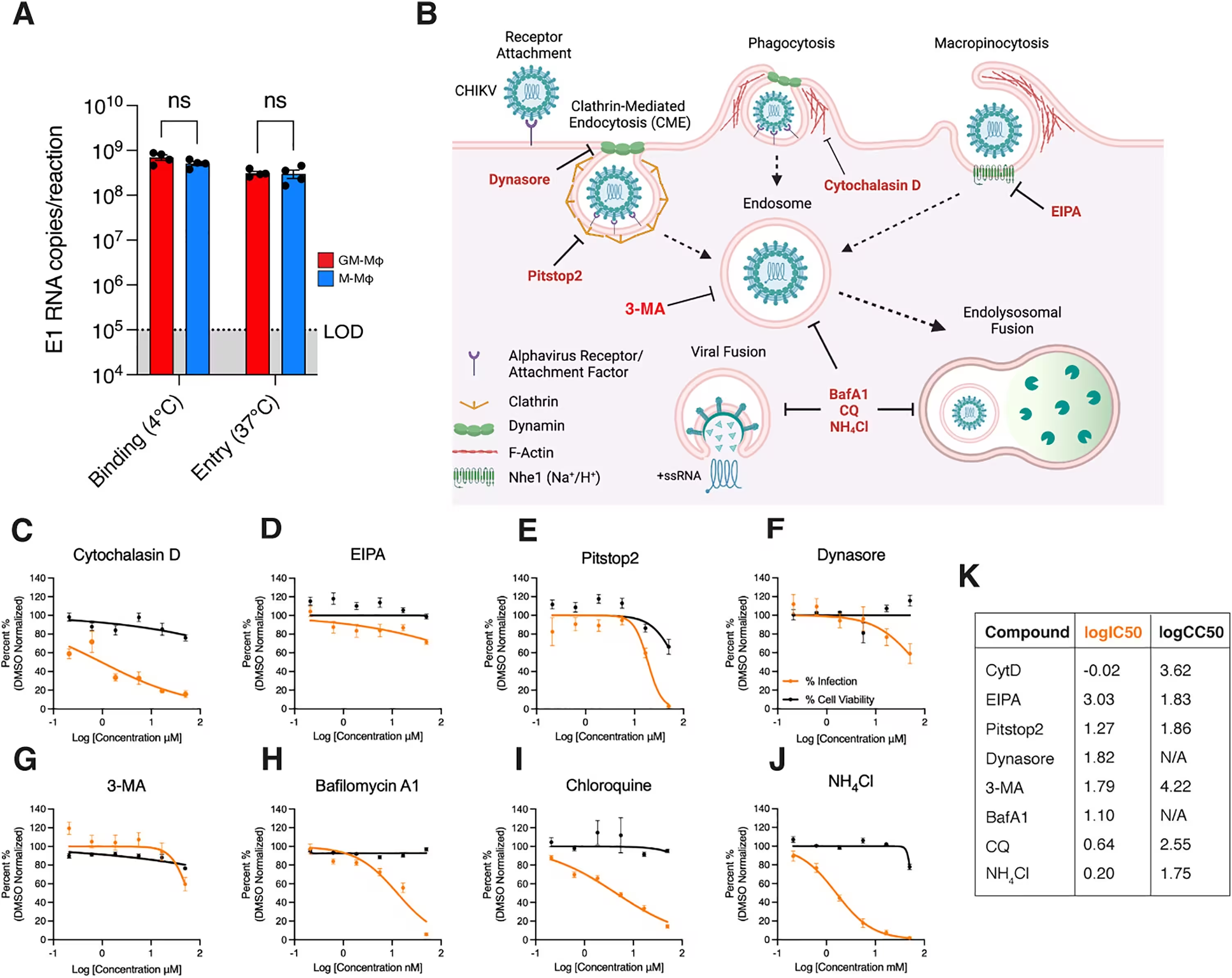
Detailed binding and entry assays revealed that both types of macrophages were equally capable of binding and internalizing Chikungunya virus particles. This indicated that the resistance observed in cells grown with macrophage colony-stimulating factor occurred after the virus entered the cell, rather than at the surface binding stage.
Chemical inhibitor tests showed that viral uptake relies on selective endocytic pathways, including clathrin- and dynamin-dependent mechanisms, along with proper endosomal acidification. However, cells grown with macrophage colony-stimulating factor mounted a much more robust antiviral response, producing significantly higher levels of alpha interferon and the chemokine IP-10.
Unraveling Interferon Suppression Mechanisms
Further genetic and molecular analyses demonstrated that Chikungunya infection actively suppressed interferon-stimulated gene expression in cells primed by granulocyte-macrophage colony-stimulating factor. This active viral suppression impaired the host defense mechanism in those specific cells, whereas interferon signaling remained fully intact in cells grown with macrophage colony-stimulating factor.
The authors suggest that these differentiation-dependent differences help explain how macrophages might serve as viral reservoirs in chronic disease. By promoting transient viral replication and blunting early interferon responses, certain immune environments could facilitate viral persistence and subsequent dissemination into joint tissues during infection.
The research was published in PLOS Pathogens on November 13, 2026.
This article has been fact checked for accuracy, with information verified against reputable sources. Learn more about us and our editorial process.
Last reviewed on .
Article history
- Latest version
Reference(s)
- Veloz, Jeury., et al. “GM-CSF and M-CSF driven differentiation differentially regulates Chikungunya virus infection and antiviral responses in human monocyte-derived macrophages.” PLOS Pathogens, vol. 22, no. 11, November 13, 2026, pp. e1014773 Public Library of Science (PLoS), doi: 10.1371/journal.ppat.1014773. <https://doi.org/10.1371/journal.ppat.1014773>.
- Posted by Faisal Qureshi