Targeted Gold Nanoparticles Outperform Standard Drug in Radiation Therapy
Engineered ultrasmall gold nanoclusters successfully amplify radiation damage in resistant breast cancer models without oxygen dependence
Tiny gold particles guided by special targeting peptides outperform a standard clinical drug at boosting radiation therapy in aggressive breast cancer models, according to a study published in PLOS One. The research investigates whether these engineered nanoclusters could overcome the notorious resistance that triple-negative breast cancer shows toward standard radiation treatments.
How Gold Nanoclusters Target Cancer Cells
Triple-negative breast cancer is a particularly aggressive form of the disease that lacks common hormonal receptors, making it especially difficult to treat effectively. Radiation therapy is a cornerstone of cancer care, but doctors must limit the radiation dose to avoid destroying healthy surrounding tissue. To solve this dilemma, researchers synthesized ultrasmall gold nanoclusters measuring just 1.88 nanometers across, decorating them with a cyclic peptide known as RGDfC. This peptide binds specifically to integrin proteins that are heavily overexpressed on the surface of breast cancer cells.
The team led by scientists at Tianjin Medical University Cancer Institute and Hospital designed these particles through a one-step biomineralization process. When exposed to X-rays, heavy metal atoms like gold absorb radiation photons and emit secondary electrons that deposit intense energy directly inside tumor cells. This physical boost amplifies DNA damage without requiring oxygen, bypassing one of the major limitations of standard radiosensitizing drugs.
Direct Comparison Against a Clinical Drug
To evaluate performance, the investigators conducted side-by-side experiments comparing the gold nanoclusters against sodium glycididazole, a small-molecule radiosensitizer widely used in clinical practice. While sodium glycididazole relies heavily on oxygen to fix radiation-induced DNA damage, tumors often suffer from severe hypoxia, or low oxygen levels, which renders such drugs less effective. The researchers tested both agents on 4T1 murine breast cancer cells under both normal oxygen conditions and chemical pseudo-hypoxia induced by cobalt chloride.
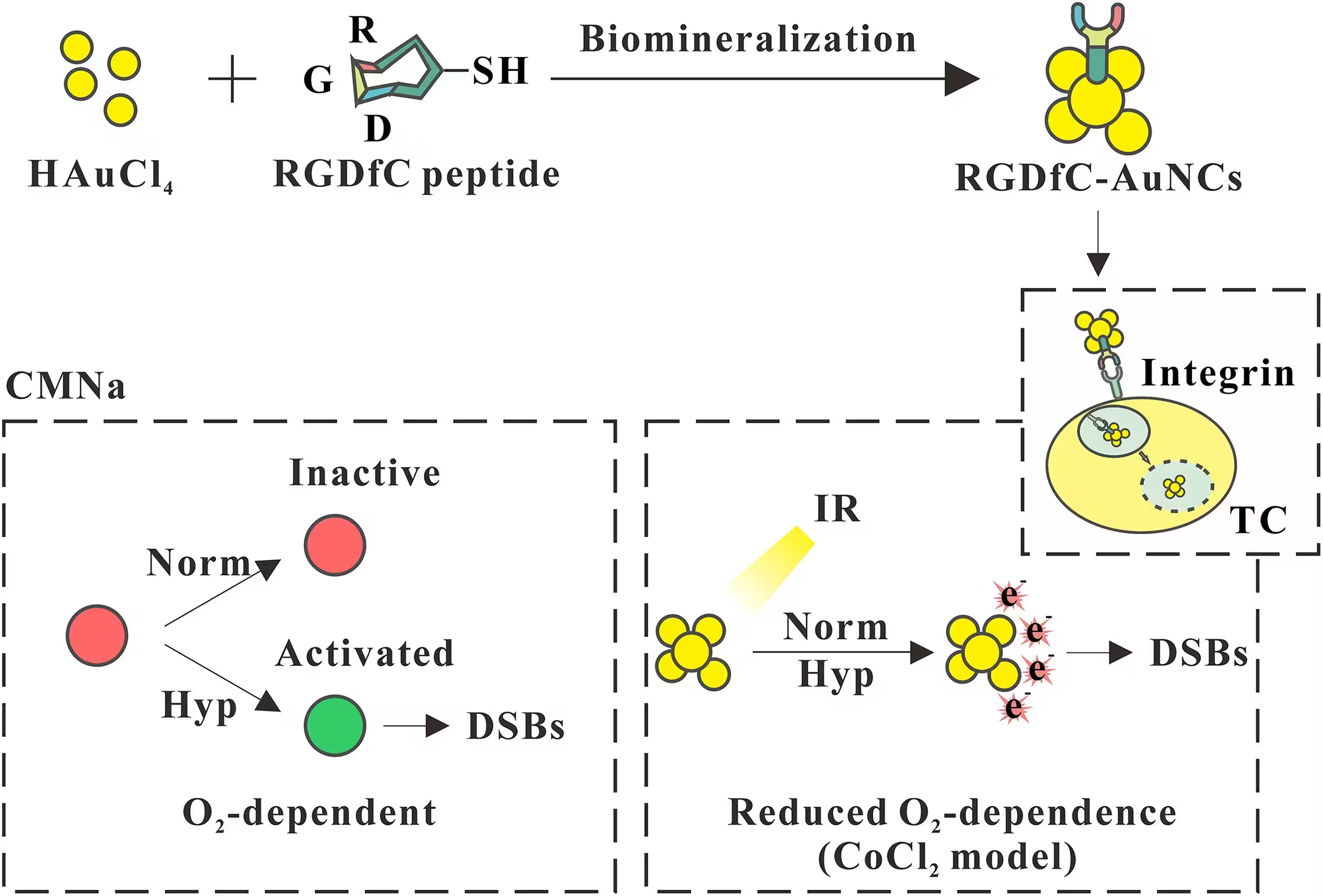
The cell viability assays revealed that the gold nanoclusters maintained strong radiosensitizing performance across both normal and low-oxygen environments. Under hypoxic conditions, the clinical drug showed modest improvement, but the gold nanoclusters consistently achieved higher radiation enhancement ratios across graded X-ray doses from two to eight gray units. The advantage of the gold nanoclusters became more pronounced over time, peaking at three days post-irradiation.
Tumor Shrinkage in Mouse Models
Following the cellular assays, the team tested the therapy in female BALB/c mice bearing subcutaneous breast cancer tumors. Animals receiving an intravenous injection of the gold nanoclusters twenty-four hours before receiving an eight-gray dose of local radiation experienced significantly greater tumor growth inhibition. The tumor growth inhibition rate reached 68.29 percent in the nanocluster group, compared to 50.61 percent for the clinical drug group and 36.12 percent for radiation alone.
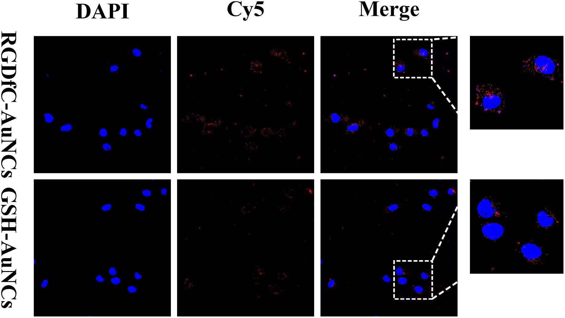
Histological examination of the excised tumor tissues confirmed these measurements. Staining with antibodies against gamma-H2AX, a well-known biomarker for double-strand DNA breaks, showed a much higher density of damaged cell nuclei in the mice treated with the targeted gold nanoclusters and radiation. Furthermore, biodistribution tracking via inductively coupled plasma mass spectrometry showed that the particles accumulated efficiently in tumor tissue while clearing progressively from the kidneys and other vital organs over seventy-two hours.
Safety Profiles and Future Directions
Short-term biosafety evaluations indicated that the gold nanoclusters caused minimal toxicity across a broad concentration range in both normal fibroblast cells and cancer cells. Laboratory tests examining mouse blood parameters, serum biochemistry, and major organ histology showed no obvious pathological lesions or inflammatory responses during the fourteen-day observation window.
Despite these promising findings, the authors note several limitations in their work. The cellular radiosensitization data relied on metabolic viability assays rather than long-term clonogenic survival assays. Additionally, the study did not perform competitive blocking experiments to definitively prove receptor-mediated active targeting, and the biosafety assessment was restricted to a two-week period. Future research will need to address long-term material clearance and explore extended treatment schedules to validate clinical potential.
The research was published in PLOS One on November 16, 2026.
This article has been fact checked for accuracy, with information verified against reputable sources. Learn more about us and our editorial process.
Last reviewed on .
Article history
- Latest version
Reference(s)
- Song, Xiangchao., et al. “RGDfC-functionalized gold nanoclusters versus the clinical radiosensitizer sodium glycididazole in triple-negative breast cancer radiotherapy.” PLOS One, vol. 21, no. 11, November 16, 2026, pp. e0362892 Public Library of Science (PLoS), doi: 10.1371/journal.pone.0362892. <https://doi.org/10.1371/journal.pone.0362892>.
Cite this page:
- Posted by Anjali Singh