Ancient Choanoflagellate Gene Reprograms Mouse Cells, Producing Mosaic Mice
Biotechnology

Ancient Choanoflagellate Gene Reprograms Mouse Cells, Producing Mosaic Mice

Ancient single‑celled organism genes reprogram mouse cells, shaping embryonic development and showing a billion‑year evolutionary link.

By Rohan Kumar
Published:
Email this Article
Scientists Inserted A Gene From Before Animal Life Existed Into Mice What Happened Next Left Them Stunned Scaled
Image credit: Gao Ya and Alvin Kin Shing Lee, with thanks to the Centre for Comparative Medicine Research (CCMR) for their support | Dungrela Publishing

Researchers have tapped a genetic mechanism that predates animal life to rewire mouse cells, steering embryonic development and yielding chimeric mammals that display distinct inherited characteristics, as reported in Nature Communications.

A Reverse Evolution Experiment

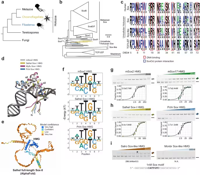
The investigation flips conventional developmental biology on its head: ancient genes, typically found in single‑celled organisms, were inserted into mouse cells and sparked a cascade of changes normally reserved for complex multicellular organisms. The team focused on choanoflagellates—microscopic eukaryotes regarded as the closest living relatives of animals—and examined their Sox‑like regulatory genes, which were once thought to be exclusive to multicellular life. When these primordial genes entered mouse cells, they did more than persist as relics; they actively remodeled cellular identity, driving cells into a pluripotent state capable of forming diverse tissue types.

41467 2024 54152 Fig1 Html
a Phylogeny of holozoans and (b) Reduced phylogenetic tree of animal and unicellular Sox. c Sequence logos representing the High Mobility Group (HMG) domain of human Sox genes, Sox-like sequences found in unicellular holozoans, and human TCF/LEF genes. Residues reported to direct DNA and protein interactions are boxed in red and blue, respectively13,28,55. d Structural Models of the Sox2 DNA binding domains (DBD) superimposed with HMG Salpingoeca helianthica (Salhel), Mylnosiga fluctuans (Myflu), and Pigoraptor chileana (Pchi). e The predicted protein structure of full-length Salhel Sox-I by AlphaFold3, with model confidence color-coded. f Energy logos derived from Spec-seq using a set of sequences with one nucleotide difference to the consensus Sox motif (CATTGTT). g–i Binding of the HMG box DBD with apparent Kd shown as mean ± SD (n = independent experiments) (g) mouse Sox2 (n = 4), and Sox17 (n = 3), (h) Salhel Sox-I (n = 3) and Pchi Sox (n = 5), and (i) Sox-like sequences from Salpingoeca rosetta (Salro) (The asterisk shows the lane with 250 nM protein which is the highest concentration used for Pchi Sox) and Monosiga brevicollis (Monbr) to consensus Sox DNA (n = 3).Credit: Nature Communications 

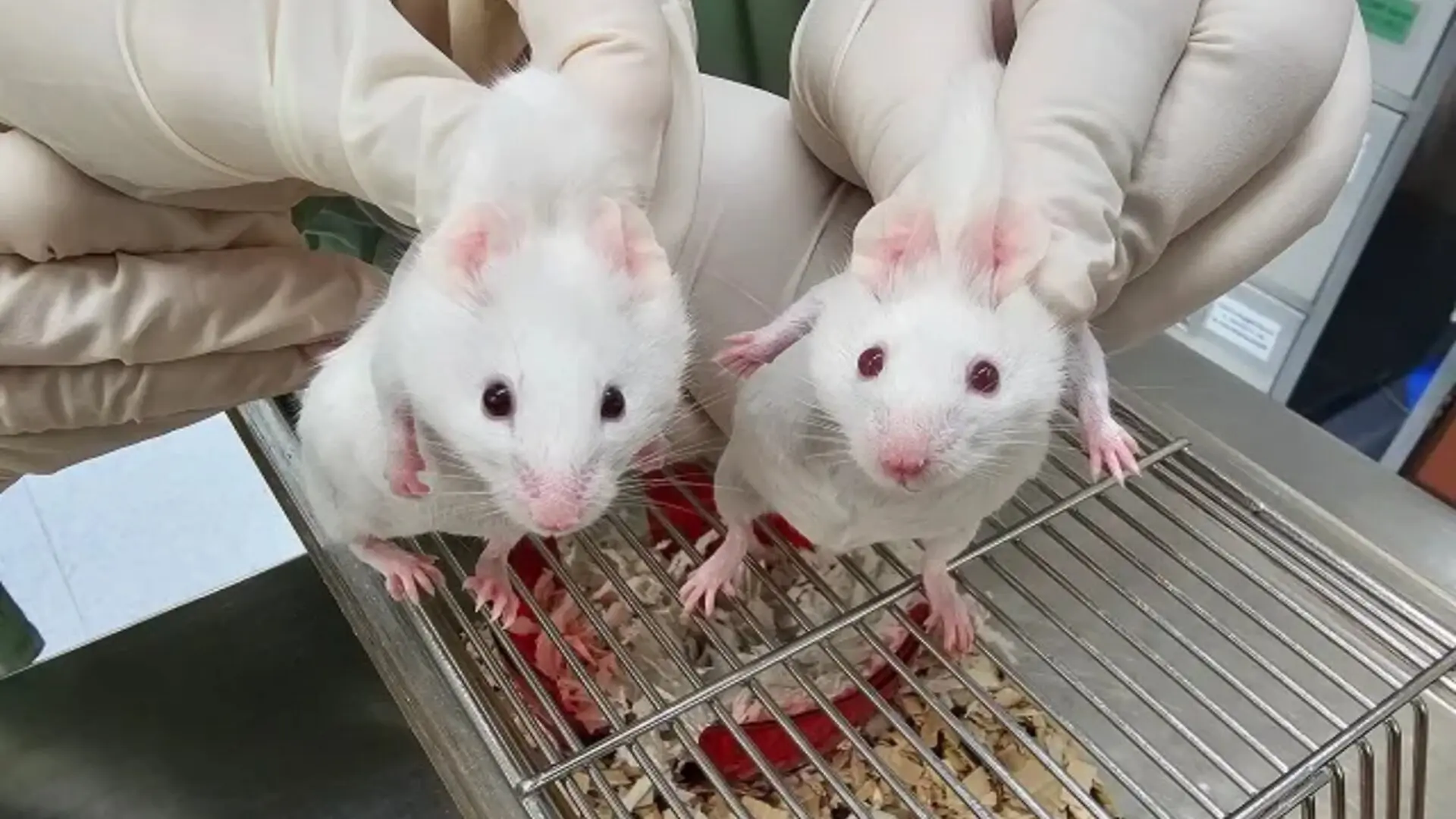
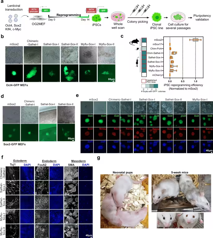
These engineered cells were coaxed into induced pluripotent stem cells that later merged with developing embryos. The resulting mice exhibited visible mosaics—black fur patches and dark eyes—signifying the integration of genetically distinct cell lines. The authors interpret the outcome as evidence that core mechanisms governing cellular flexibility have been conserved for nearly a billion years, challenging prevailing ideas about the timing of developmental gene emergence.

Re‑engineering Stem‑Cell Pathways with Ancient Genes

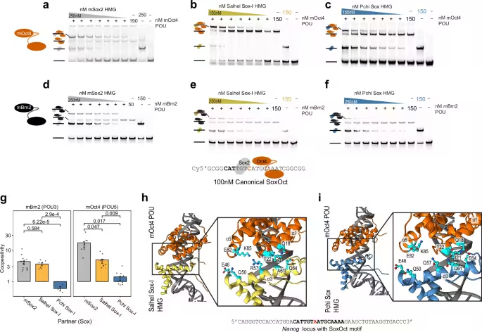
In the reported study (Nature Communications), scientists swapped a pivotal mammalian stem‑cell regulator for a choanoflagellate counterpart. Although choanoflagellates lack true stem cells, they possess Sox‑like genes previously believed to be animal‑specific. When these genes entered mouse fibroblasts, they ignited pluripotency circuits that normally rely on mammalian transcription factors. The induced pluripotent stem cells were subsequently introduced into early embryos, where they contributed to multiple lineages.

Mosaic animals emerged, confirming that the ancient genes exerted functional influence throughout development. The authors highlight the surprising compatibility between distant evolutionary lineages, suggesting that modern developmental processes still lean on deeply conserved molecular scaffolds.

41467 2024 54152 Fig2 Html
a Schematic illustration of the procedure of mouse induced pluripotent stem cell (iPSC) reprogramming from mouse embryonic fibroblasts (MEFs) carrying an Oct4-GFP reporter (OG2MEFs) and the establishment of clonal iPSC line for pluripotency validation. b Representative microscope images show iPSC colonies generated by mSox2 and Sox factors of Choanoflagellates on reprogramming day 14. Scale bar, 80μm. Chimeric-Salhel-I (Chimeric-Salhel-Sox-I), HMG of Salhel-Sox-I fused with mSox2 NTD and CTD; Salhel-Sox-I, full-length Salhel Sox-I; Salhel-Sox-II, full-length Salhel Sox-II; Myflu-Sox-I, full-length Myflu Sox-I; Myflu-Sox-II, full-length Myflu Sox-II. c Quantification of iPSC reprogramming efficiency by Sox variants. The heatmap depicts the number of experiments with observation of GFP-positive colonies, with the red frame highlighting the ability of the variants to produce iPSCs with confirmed pluripotency through the establishment of stable clonal iPSC lines. Chim-Salhel-I, Chimeric-Salhel-Sox-I; Chim-Pchi, Chimeric-Pchi-Sox. The box plot shows the reprogramming efficiency of Sox variants normalized by the number of iPSC colonies generated by mSox2. (n = 7 technical replicates in total, 2 biological replicates each with 2 technical replicates and 1 biological replicates including 3 technical replicates). The box displays the interquartile range, with the left edge representing the lower quartile (25th percentile) and the right edge indicating the upper quartile (75th percentile). The median value is shown as a line splitting the box. The silhouettes of the species are sourced from PhyloPic (http://phylopic.org). d Representative images of iPSC colonies derived from MEFs carrying a Sox2‑GFP reporter on reprogramming day 14. Scale bar, 80 μm. e Expression of pluripotency markers of clonal iPSC lines derived by choanoflagellate Sox examined by immunocytochemistry staining. Scale bar, 40μm. f Immunocytochemistry of differentiated iPSC lines stained for markers of the 3 germ layers: Class III beta‑tubulin (Tuj1), Forkhead box protein A2 (FoxA2), α‑smooth muscle actin (SMA). Scale bar, 40 μm. g Chimeric mice generated from full-length Salhel‑Sox‑I iPSC lines displaying black coat patches and eyes (indicated by arrows) representing their iPSC origin, in contrast to the wildtype mouse exhibiting a white coat and red eyes.Credit: Nature Communications 

Reflecting on the work, study author Dr Alex de Mendoza noted,

“By successfully creating a mouse using molecular tools derived from our single‑celled relatives, we’re witnessing an extraordinary continuity of function across nearly a billion years of evolution.”

The authors also propose that genes linked to stem‑cell behavior may have arisen before stem cells themselves, prompting a rethink of early genetic regulation.

Implications for the Origin of Cellular Plasticity

Beyond the technical breakthrough, the study raises fundamental questions about the emergence of biological complexity. Choanoflagellates lack true stem cells, yet they harbor genetic elements that mirror those used in animal development. This paradox hints that molecular systems associated with multicellular organization may have originated in a unicellular context and later been repurposed as organisms grew more complex. As Dr de Mendoza explained,

“Choanoflagellates don’t have stem cells, they’re single‑celled organisms, but they have these genes, likely to control basic cellular processes that multicellular animals probably later repurposed for building complex bodies,”

The presence of such genes in single‑celled life supports the idea that evolutionary innovation often stems from the reuse of existing components rather than from de novo invention. Cellular identity, differentiation, and developmental control may therefore rest on ancient regulatory frameworks that predate animals by hundreds of millions of years. This perspective shifts focus from abrupt evolutionary leaps toward gradual adaptation of long‑standing molecular architectures, blurring the genetic boundary between unicellular and multicellular organisms.

41467 2024 54152 Fig3 Html
a–f Heterodimer EMSAs with 50 or 100 nM Cy5 labeled canonical SoxOct DNA elements to monitor the heterodimer formation of POU factors (a–c) 150–190 nM mOct4 or (d–f) 50 nM mBrn2 with Sox factors – (a–d) mSox2 (a–e) Salhel Sox‑I or (c–f) Pchi Sox HMG. POU factors are kept at a constant concentration indicated by + signs, triangles indicate different concentrations of Sox with the highest concentration indicated, and – sign indicates absences of either Sox or POU or both for controls. g Quantifications of heterodimer EMSAs and calculation of cooperativity factors according to (Ng et al. 2012) with the y axis depicted in log10 scale (mean ± SEM) with n = independent experiments. Oc4/Sox2 (n = 4), Oc4/Pchi Sox (n = 3), Oct4/Salhel Sox‑I (n = 4) and Brn2 with Sox2, Pchi Sox and Salhel Sox‑I (n = 3). Adjusted p-values are shown and were determined from a Games‑Howell test with a 0.95 confidence interval after Bartlett test of homogeneity for each dataset (mOct4 – p = 2.06E‑08, mBrn2 p = 0.003637) and Kruskal‑Wallis test(One‑way). h, i Structural models of heterodimer complexes on canonical SoxOct motifs of (f) Salhel Sox‑I HMG‑mOct4 POU complex or (g) Pchi Sox HMG‑mOct4 POU complex highlighting differences at the heterodimer interface (i.e. positions 57, 61 and 64 previously predicted to impact dimer formation).Credit: Nature Communications 

Chimera Formation and Developmental Flexibility

One of the most striking visual outcomes was the generation of chimeric mice—organisms composed of cells from distinct genetic origins. After injecting the engineered stem cells into embryos, they integrated across multiple tissues, producing observable mosaics in coat and eye coloration that served as biological markers of cellular contribution. The stability of these reprogrammed cells within a living organism demonstrates that pluripotent states can be sustained beyond the early embryo.

The authors note that such compatibility underscores the robustness of developmental systems, which can accommodate genetically diverse inputs without compromising overall growth. Dr Ralf Jauch added,

“Studying the ancient roots of these genetic tools lets us innovate with a clearer view of how pluripotency mechanisms can be tweaked or optimised,”

Overall, the work suggests that developmental plasticity is a deeply rooted biological trait, extending far beyond the origins of animals and offering new avenues for biomedical research.

Fact Checked

This article has been fact checked for accuracy, with information verified against reputable sources. Learn more about us and our editorial process.

Last reviewed on .

Article history

  • Latest version

Reference(s)

  1. Gao, Ya. “The emergence of Sox and POU transcription factors predates the origins of animal stem cells - Nature Communications.”, vol. 15, no. 1, November 14, 2024, pp. 9868 Nature, doi: 10.1038/s41467-024-54152-x. <https://www.nature.com/articles/s41467-024-54152-x>.

Cite this page:

Kumar, Rohan. “Ancient Choanoflagellate Gene Reprograms Mouse Cells, Producing Mosaic Mice.” BioScience. BioScience ISSN 2521-5760, 29 June 2026. <https://www.bioscience.com.pk/en/subject/biotechnology/scientists-inserted-a-gene-from-before-animal-life-existed-into-mice-what-happened-next-left-them-stunned>. Kumar, R. (2026, June 29). “Ancient Choanoflagellate Gene Reprograms Mouse Cells, Producing Mosaic Mice.” BioScience. ISSN 2521-5760. Retrieved June 29, 2026 from https://www.bioscience.com.pk/en/subject/biotechnology/scientists-inserted-a-gene-from-before-animal-life-existed-into-mice-what-happened-next-left-them-stunned Kumar, Rohan. “Ancient Choanoflagellate Gene Reprograms Mouse Cells, Producing Mosaic Mice.” BioScience. ISSN 2521-5760. https://www.bioscience.com.pk/en/subject/biotechnology/scientists-inserted-a-gene-from-before-animal-life-existed-into-mice-what-happened-next-left-them-stunned (accessed June 29, 2026).

Follow us on social media

End of the article