Choline Supplement Rejuvenates Mitochondria By Restoring Key Lipid, New Study Shows
A newly identified molecule that wanes with age disrupts cellular energy networks, but dietary boosting can revive function in older organisms.
Long‑standing research has shown that mitochondria—the cellular power stations—gradually lose function as organisms age, yet the underlying cause remained elusive. A recent paper in Nature Communications identifies a steady reduction in the membrane lipid phosphatidylcholine as a key driver of this decline.
The investigation, led by Dr Maria Ermolaeva at the Leibniz Institute on Aging in Jena, combined work in the nematode Caenorhabditis elegans with analyses of human cell lines and extensive clinical datasets. Their results imply that certain aspects of mitochondrial senescence are not immutable and could be amenable to targeted correction.
Failure of Cellular Power Grids
Beyond ATP production, mitochondria form an adaptable web inside cells, allowing them to exchange metabolites, repair damage, and meet shifting energy needs. This flexibility, known as metabolic plasticity, relies heavily on mitochondrial fusion, a process that merges smaller organelles into larger, more robust networks.
Phosphatidylcholine, one of the most abundant lipids in mitochondrial membranes, is essential for maintaining membrane fluidity and enabling fusion events. The authors report that levels of this lipid naturally decline with age, leading to increasingly fragmented and less efficient mitochondrial networks, as described in the Nature Communications study.
Ermolaeva likens the situation to an aging electrical grid: “Connections weaken and currents lag, so although energy production persists, it becomes less efficient and less adaptable to demand,” she explained. This loss of flexibility mirrors age‑related disorders such as diabetes, where cells struggle to meet rapid energy fluctuations.
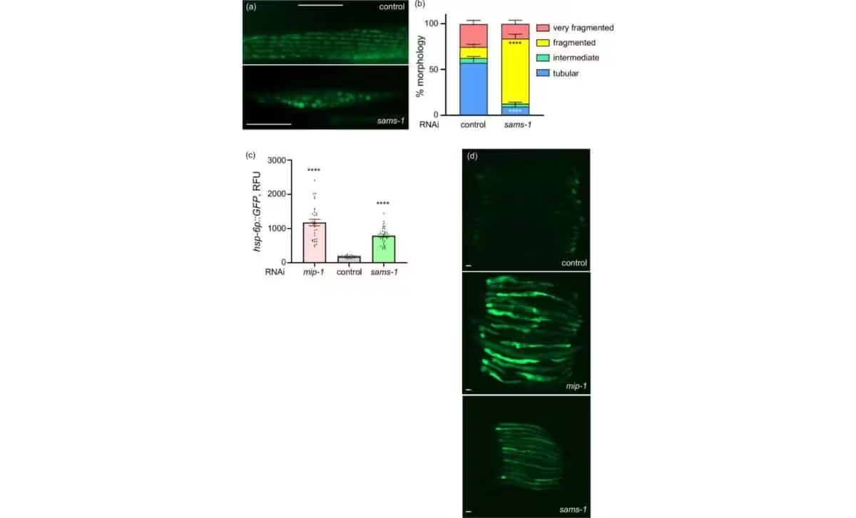
When the team knocked out genes responsible for phosphatidylcholine synthesis in young worms, their mitochondria rapidly adopted an aged, fragmented appearance. Remarkably, feeding the animals a phosphatidylcholine‑rich diet restored network integrity within just two days.
Human Insights Reveal a Post‑Menopausal Lipid Dip
To translate the worm findings to humans, the researchers examined transcriptomic data from the GTEx Project and metabolomic profiles from the UK Biobank, which together encompass hundreds of thousands of individuals.
Across multiple tissues, the expression of the enzyme that generates phosphatidylcholine declines with age, especially in lipid‑rich organs like adipose tissue. Blood concentrations of the lipid drop steadily in men, while women experience a sharper decrease around menopause—a stage already linked to weakened mitochondrial performance and heightened fatigue.
Higher phosphatidylcholine levels were associated with lower circulating lactate—a marker of mitochondrial stress—as well as improved gait speed and memory scores. Conversely, reduced levels correlated with a greater burden of comorbidities and an unfavorable metabolic profile.
Crucially, the benefits of supplementation persisted even when introduced later in life. Supplying choline—a dietary precursor to phosphatidylcholine—to aging worms enhanced both mitochondrial architecture and respiration. In cultured human cells, choline protected against metformin‑induced mitochondrial stress.
“Our results demonstrate that mitochondrial and systemic aging are at least partly reversible,” Ermolaeva said. “Understanding the mechanisms opens the door to targeted interventions.”
The authors caution that human trials are required before clinical recommendations can be made. Variables such as gut microbiome composition may influence how choline and phosphatidylcholine are processed in people, potentially affecting outcomes compared with laboratory models. Nevertheless, the study shifts the narrative from inevitability toward the prospect of fine‑tuning cellular aging.
This article has been fact checked for accuracy, with information verified against reputable sources. Learn more about us and our editorial process.
Last reviewed on .
Article history
- Latest version
Reference(s)
- Poliezhaieva, Tetiana. “Aging-associated decline of phosphatidylcholine synthesis is a malleable trigger of natural mitochondrial aging - Nature Communications.”, vol. 17, no. 1, April 18, 2026, pp. 3589 Nature, doi: 10.1038/s41467-026-71508-7. <https://www.nature.com/articles/s41467-026-71508-7>.
Cite this page:
- Posted by Rohan Kumar