AI Predicts Cellular RNA and Proteins From Routine Hospital Slides
A new deep learning framework leverages biological gene relationships to map molecular profiles from standard pathology tissue stains.
A new artificial intelligence framework successfully reconstructs thousands of hidden cellular molecular profiles directly from routine hospital microscope slides. By analyzing standard tissue sections stained with the pink and purple dyes common in pathology labs, the model predicts both RNA transcript activity and protein distributions at single-cell resolution. Researchers developed the system to unlock molecular insights from massive archives of historical patient samples without requiring expensive, specialized spatial assays.
Bridging RNA and Protein Data Across Unpaired Tissues
Obtaining detailed maps of gene expression and protein markers across a tissue sample usually demands technically demanding techniques like in situ spatial transcriptomics or multiplex immunofluorescence. Because these advanced assays are costly, laboratories rarely perform both RNA and protein measurements on the exact same tissue section. The research team behind the new model, published on the arXiv preprint server, sought to overcome this hurdle by creating a unified system called SCOPE, which stands for Single-Cell Omics Prediction via RNA-protein prior Enhancement.
Led by computer scientists and bioinformaticians from Harvard Medical School, Stanford University, and the Beijing University of Civil Engineering and Architecture, the authors designed their approach around biological connections. Although RNA molecules and proteins represent distinct molecular types, the genes that encode them share a natural biological link. The framework exploits this correspondence to allow supervision from either RNA data or protein data to strengthen a shared gene-level representation.
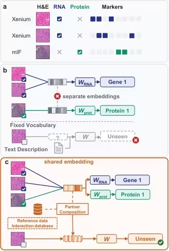
Combining Knowledge Bases and Dual-Resolution Architecture
To predict genes and markers that lack direct supervision during training, the authors incorporated biological knowledge graphs from databases like STRING and Reactome alongside single-cell co-expression patterns. This biomarker co-encoding strategy allows the algorithm to transfer knowledge from well-studied partner genes to unseen targets. The system reads hematoxylin and eosin (H&E) images using a dual-branch architecture that evaluates data at two different scales. A cell-level branch scores individual cell nuclei based on frozen representations from a pathology foundation model called H-optimus-1, while a dense expression field branch maps molecular abundance across individual pixels.
The researchers trained their framework on extensive datasets comprising 43 Xenium spatial transcriptomics sections spanning 10 organs and 76 CODEX multiplexed imaging sections from three donors. This training yielded approximately 7.87 million Xenium cells and 26.49 million CODEX cells. To evaluate generalizability, the team tested the model on 16 independent Xenium sections from 16 organs, including six organs completely absent from the training data, as well as 22 CODEX sections from a separate donor and two large external colorectal cancer patient cohorts containing millions of cells.
Performance Gains Across Multiple Biological Tissues
According to the evaluation metrics reported by the authors, the unified framework outperformed existing baseline models across diverse test sets. On the 16 test sections covering 10 organs, the system achieved an average Pearson correlation of 0.484 on the ten genes with the highest variance per section, surpassing the strongest fine-tuned baseline. Across external protein cohorts, the method improved average correlation for seen protein markers and successfully generalized to predict unseen protein markers that were entirely absent from training supervision.
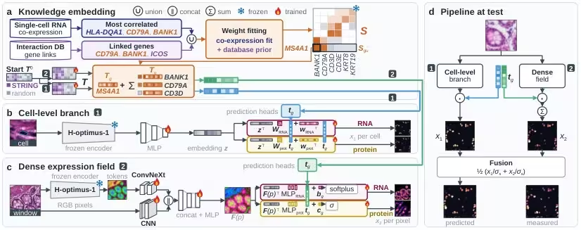
The ablation studies conducted by the research team shed light on why the joint training approach succeeded. When the model was trained on both modalities simultaneously, protein prediction accuracy improved even though the training sections did not measure both RNA and protein together. Furthermore, equal-weight late fusion of the cell-level branch and the dense pixel-level branch consistently surpassed the performance of either individual branch alone across test settings.
Limitations and Future Path for Pathology AI
Despite these advances, the study authors noted certain limitations inherent to predicting molecular profiles from standard histology. The accuracy of the predictions depends heavily on the quality of nuclear segmentation and the biological relationships captured in the underlying interaction databases. Unseen markers lacking direct supervision still rely heavily on the fidelity of inferred gene co-expression weights and prior knowledge networks. Future work will likely focus on expanding validation across an even wider array of rare pathologies and incorporating larger multimodal foundation models to refine spatial resolution and clinical applicability.
The study was posted to the arXiv preprint server on November 18, 2026.
This article has been fact checked for accuracy, with information verified against reputable sources. Learn more about us and our editorial process.
Last reviewed on .
Article history
- Latest version
Reference(s)
- Tao, Leyang. “SCOPE: Single-Cell Omics Prediction from Histology via RNA-Protein Prior Enhancement.” arXiv.org <http://arxiv.org/abs/2611.24104v1>.
Cite this page:
- Posted by Hassan Raza