Enzyme Deletion Prevents Fatal Lung Bleeding and Boosts Repair
Biology

Enzyme Deletion Prevents Fatal Lung Bleeding and Boosts Repair

Removing a key immune kinase reprograms regulatory T cells to heal damaged lung barriers and suppress systemic inflammation.
By Hassan Raza
Published:
Email this Article
Enzyme Deletion Prevents Fatal Lung Bleeding and Boosts Repair
Photo by Google DeepMind on Unsplash

Removing a single immune enzyme prevents life-threatening lung bleeding in mice and reprograms specialized guardian cells to repair damaged tissue, a study reveals.

Pulmonary hemorrhage is a severe and often fatal complication of autoimmune diseases where the delicate barrier between air sacs and blood vessels in the lungs breaks down. Traditional treatments rely on heavy immunosuppression, yet mortality rates remain stubbornly high. Now, researchers have identified a potential molecular switch that not only halts this destructive inflammation, but actively drives tissue repair. The findings were published in eLife by Mohammad Saddam Hossain and colleagues.

How Eliminating an Enzyme Protects the Lungs

The team focused on interleukin-2-inducible T-cell kinase, an enzyme known to amplify immune responses and drive inflammation. By testing mice genetically lacking this enzyme, the researchers discovered that the animals were nearly completely protected against chemically induced pulmonary hemorrhage. While untreated control animals suffered massive blood accumulation in their lungs and lost architectural integrity, the modified mice maintained healthy tissue structures and showed greatly improved survival.
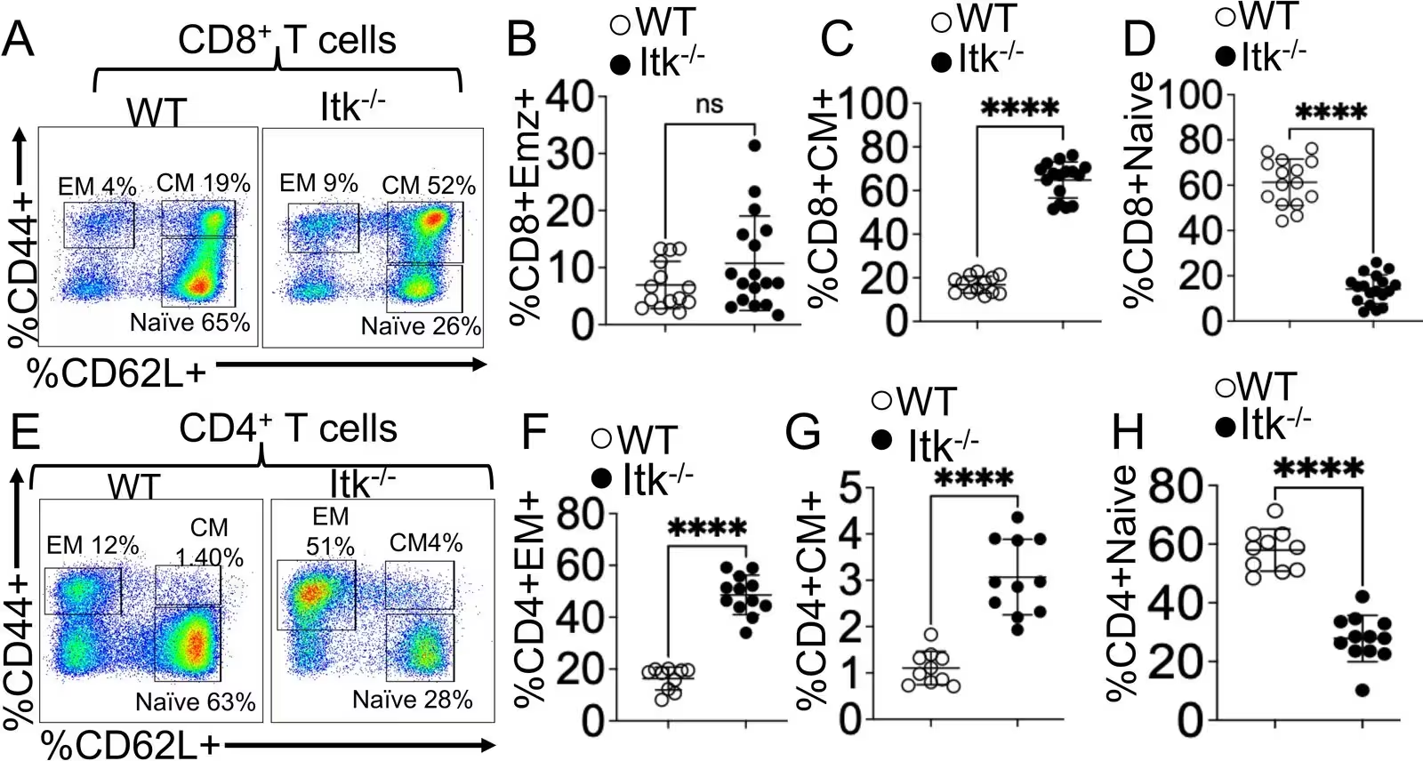
Figure 1: ITK deficiency promotes a predominantly memory-like T-cell phenotype. (A–D) Analysis of the CD8+ T-cell compartment in WT and Itk⁻/⁻ mice. Freshly isolated splenocytes were gated on CD3+ lymphocytes and further subdivided into CD8+ and CD4+ T-cell populations. (A) Representative flow cytometry plots showing CD44 and CD62L expression within CD8+ T cells. (B–D) Quantification of effector memory (EM; CD44+CD62L⁻), central memory (CM; CD44+CD62L+), and naive (CD44⁻CD62L+) CD8+ T-cell subsets. (E–H) Analysis of the CD4+ T-cell compartment. (E) Representative flow cytometry plots showing CD44 and CD62L expression within CD4+ T cells. (F–H) Quantification of EM, CM, and naive CD4+ T-cell frequencies. Data are presented as mean ± SEM (n = 10–15 mice per group). Results are representative of six independent experiments. Statistical significance was determined by an unpaired two-tailed Student’s t-test. ****p ≤ 0.0001; ns, not significant (p > 0.05).
ITK deficiency promotes a predominantly memory-like T-cell phenotype. (A–D) Analysis of the CD8+ T-cell compartment in WT and Itk⁻/⁻ mice. Freshly isolated splenocytes were gated on CD3+ lymphocytes and further subdivided into CD8+ and CD4+ T-cell populations. (A) Representative flow cytometry plots showing CD44 and CD62L expression within CD8+ T cells. (B–D) Quantification of effector memory (EM; CD44+CD62L⁻), central memory (CM; CD44+CD62L+), and naive (CD44⁻CD62L+) CD8+ T-cell subsets. (E–H) Analysis of the CD4+ T-cell compartment. (E) Representative flow cytometry plots showing CD44 and CD62L expression within CD4+ T cells. (F–H) Quantification of EM, CM, and naive CD4+ T-cell frequencies. Data are presented as mean ± SEM (n = 10–15 mice per group). Results are representative of six independent experiments. Statistical significance was determined by an unpaired two-tailed Student’s t-test. ****p ≤ 0.0001; ns, not significant (p > 0.05). Credit: Mohammad Saddam Hossain et al. / eLife

Further analysis revealed that the absence of the enzyme dramatically reshaped the animals’ immune landscapes. The lungs of the protected mice showed significantly fewer destructive inflammatory monocytes and neutrophils. Additionally, systemic inflammation dropped sharply, with circulating levels of harmful inflammatory cytokines like interferon-gamma, tumor necrosis factor-alpha, interleukin-6, and interleukin-17 all registering much lower.

Why Guardian T Cells Shift Into Repair Mode

The protective effect was not merely a passive lack of inflammation, but an active shift toward healing driven by regulatory T cells, specialized white blood cells that maintain immune tolerance. Mice lacking the enzyme exhibited a marked expansion of these regulatory populations in both the spleen and the lungs. When these specialized cells were isolated and transferred into diseased wild-type mice, they successfully rescued the animals from lung hemorrhage and restored baseline renal function.
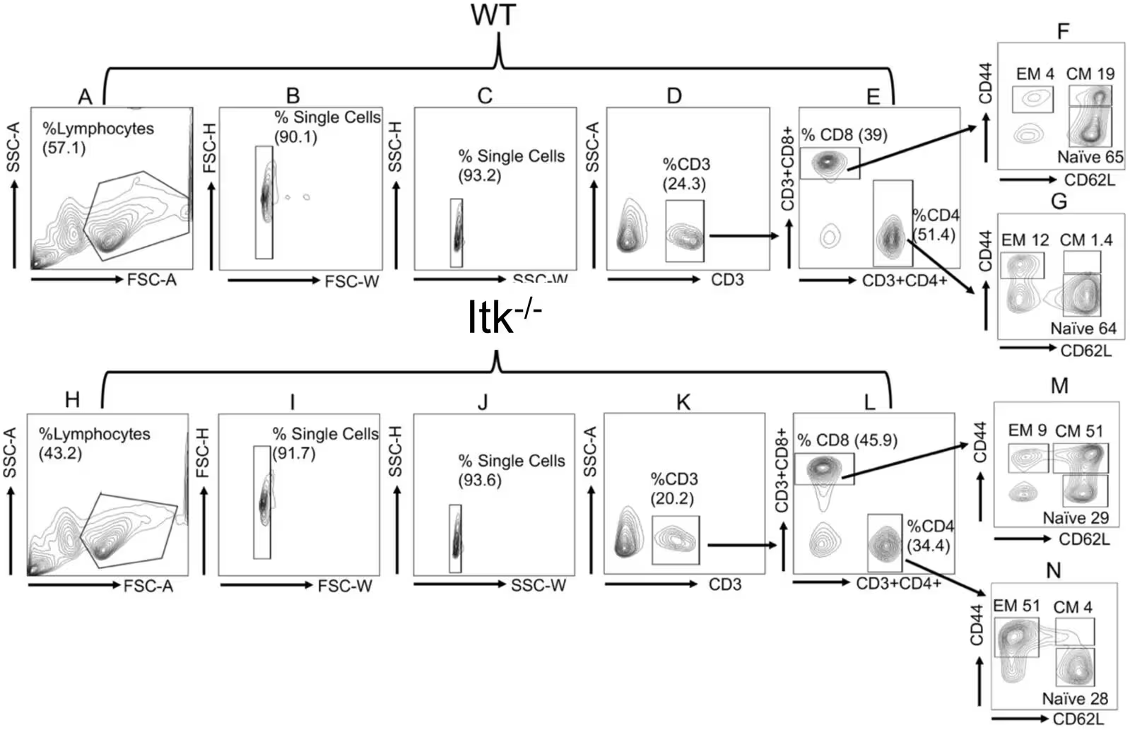
Figure 1—figure supplement 1: Gating strategies for effector and central memory phenotypes. Freshly isolated splenocytes from WT (A–G) and Itk⁻/⁻ (H–N) mice were analyzed by flow cytometry. (A, H) Lymphocyte populations were initially gated based on side scatter area (SSC-A) versus forward scatter area (FSC-A). (B, I) Lymphocytes were then gated for single cells using forward scatter height (FSC-H) versus forward scatter width (FSC-W) to discriminate doublets. (C, J) Additional singlet gating was performed using side scatter height (SSC-H) versus side scatter width (SSC-W) to exclude any remaining doublets. (D, K) CD3+ T lymphocytes were identified from the single-cell population based on SSC-A versus CD3 expression. (E, L) The CD3+ compartment was further subdivided into CD8+ and CD4+ T-cell populations. (F, M) CD8+ and (G, N) CD4+ T cells were then analyzed to identify and quantify effector memory (EM; CD44+CD62L⁻), central memory (CM; CD44+CD62L+), and naive (CD44⁻CD62L+) subsets, as depicted in the contour plot
Gating strategies for effector and central memory phenotypes. Freshly isolated splenocytes from WT (A–G) and Itk⁻/⁻ (H–N) mice were analyzed by flow cytometry. (A, H) Lymphocyte populations were initially gated based on side scatter area (SSC-A) versus forward scatter area (FSC-A). (B, I) Lymphocytes were then gated for single cells using forward scatter height (FSC-H) versus forward scatter width (FSC-W) to discriminate doublets. (C, J) Additional singlet gating was performed using side scatter height (SSC-H) versus side scatter width (SSC-W) to exclude any remaining doublets. (D, K) CD3+ T lymphocytes were identified from the single-cell population based on SSC-A versus CD3 expression. (E, L) The CD3+ compartment was further subdivided into CD8+ and CD4+ T-cell populations. (F, M) CD8+ and (G, N) CD4+ T cells were then analyzed to identify and quantify effector memory (EM; CD44+CD62L⁻), central memory (CM; CD44+CD62L+), and naive (CD44⁻CD62L+) subsets, as depicted in the contour plot Credit: Mohammad Saddam Hossain et al. / eLife

Transcriptomic sequencing revealed that removing the enzyme reprogrammed the regulatory cells to be metabolically and functionally enhanced. These enhanced guardian cells expressed significantly higher levels of amphiregulin, a growth factor protein vital for tissue repair and barrier restoration. At the same time, the cells boosted production of anti-inflammatory signaling molecules like interleukin-10, creating a robust healing environment.

What These Discoveries Mean for Future Therapies

While the study demonstrates remarkable protection in a murine model, researchers note that experiments were conducted using germline-deficient animals and cells isolated from unchallenged subjects. Additional investigations using conditional genetic models will be necessary to pinpoint the exact cell-intrinsic pathways at play. Nevertheless, the work points to the enzyme as a promising therapeutic target for treating severe inflammatory lung conditions and systemic autoimmunity without shutting down the entire immune system.

The research was published in eLife on November 17, 2026.

Fact Checked

This article has been fact checked for accuracy, with information verified against reputable sources. Learn more about us and our editorial process.

Last reviewed on .

Article history

  • Latest version

Reference(s)

  1. Hossain, Mohammad Saddam., et al. “ITK deficiency protects against autoimmune pulmonary hemorrhage in mice by promoting amphiregulin-producing regulatory T cells.” eLife, vol. 15, November 17, 2026 eLife Sciences Publications, Ltd, doi: 10.7554/eLife.111915. <https://doi.org/10.7554/eLife.111915>.

Cite this page:

Raza, Hassan. “Enzyme Deletion Prevents Fatal Lung Bleeding and Boosts Repair.” BioScience. BioScience ISSN 2521-5760, 19 November 2026. <https://www.bioscience.com.pk/en/subject/biology/itk-deficiency-protects-against-autoimmune-pulmonary-hemorrhage-in-mice-by-promoting-amphiregulin-producing-regulatory-t-cells>. Raza, H. (2026, November 19). “Enzyme Deletion Prevents Fatal Lung Bleeding and Boosts Repair.” BioScience. ISSN 2521-5760. Retrieved November 19, 2026 from https://www.bioscience.com.pk/en/subject/biology/itk-deficiency-protects-against-autoimmune-pulmonary-hemorrhage-in-mice-by-promoting-amphiregulin-producing-regulatory-t-cells Raza, Hassan. “Enzyme Deletion Prevents Fatal Lung Bleeding and Boosts Repair.” BioScience. ISSN 2521-5760. https://www.bioscience.com.pk/en/subject/biology/itk-deficiency-protects-against-autoimmune-pulmonary-hemorrhage-in-mice-by-promoting-amphiregulin-producing-regulatory-t-cells (accessed November 19, 2026).
End of the article