Computer Simulations Reveal How CYFIP1 Grips eIF4E in Cells
Advanced molecular modeling and artificial intelligence uncover a surprisingly plastic and multi-pronged interface between a key translation regulator and its cellular partner.
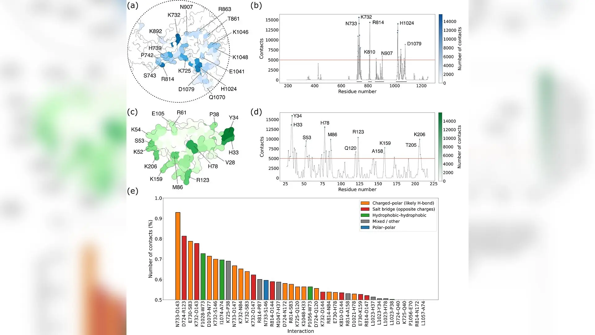
Eukaryotic initiation factor 4E, a master regulator of protein synthesis also known as eIF4E, binds to its cellular partner CYFIP1 through a surprisingly flexible and multi-pronged interface rather than a single fixed pose. The scaffold protein CYFIP1 acts as a molecular switch in human cells, oscillating between controlling the cytoskeleton and repressing messenger RNA translation by interacting with eIF4E. Dysregulation of this precise molecular handshake is heavily implicated in neurodevelopmental conditions such as autism spectrum disorder and schizophrenia, as well as various forms of cancer. Previous biochemical studies established that a short helical motif within CYFIP1 is essential for binding, but scientists lacked a complete atomic-resolution picture of how the full-length proteins associate dynamically in solution.
Mapping the Molecular Dance Through 100 Microseconds of Simulation
To uncover how these large molecules interact, a team of researchers combined all-atom molecular dynamics, data-driven docking, and coarse-grained enhanced sampling to map the structural landscape. The scientists utilized the HADDOCK3 platform alongside 10 independent replicas of funnel-restrained Temperature-Accelerated Molecular Dynamics, accumulating 100 microseconds of total sampling time. Because direct clustering of such massive, highly flexible trajectories typically collapses into poorly resolved groups, the authors trained a deep learning architecture on contact distance matrices. As detailed in a study published on the arXiv preprint server, this approach leveraged a beta-Variational Autoencoder to compress high-dimensional structural data into a two-dimensional latent space, successfully identifying distinct, energetically favorable binding conformations.
Why the Classic Binding Blueprint Falls Short
The resulting free-energy landscapes reveal that the interaction is not governed by a rigid, singular lock-and-key mechanism. Instead, the complex samples multiple distinct binding modes, categorized into three major structural clusters where CYFIP1 engages eIF4E from different angles. While the canonical helical motif acts as a primary contact hotspot in all major configurations, the simulations demonstrate that classic 4E-binding protein interfaces on eIF4E are largely bypassed. Instead, CYFIP1 heavily recruits alternative contact regions, including its Dimerization/Interaction Module and a basic patch containing specific residues like Lys810 and Arg814. These auxiliary contact points offer a compelling structural explanation for experimental findings showing that mutating the main helical motif alone only partially abolishes binding.
Connecting Translational Repression to Cytoskeletal Control
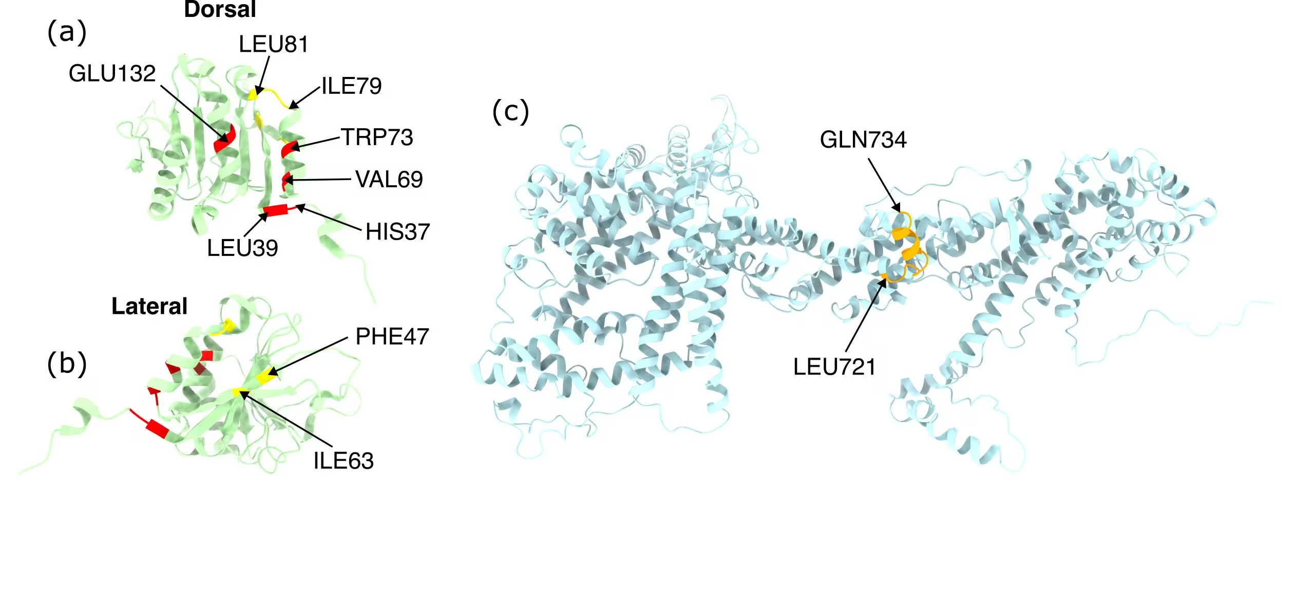
These findings point to a deeper mechanistic link between protein synthesis and actin remodeling within the cell. The region of CYFIP1 that engages eIF4E through its auxiliary contact domains overlaps with the segment responsible for protein dimerization within the WAVE Regulatory Complex, which governs actin dynamics. This structural overlap suggests that translational machinery and cytoskeletal components might compete for the same molecular surfaces. Such spatial competition provides fresh insight into how single mutations in CYFIP1 can trigger sweeping downstream effects across multiple cellular pathways.
New Blueprints for Targeted Peptide Therapies
By defining this heterogeneous ensemble of binding modes rather than a single static conformation, the work establishes a robust foundation for future therapeutic interventions. Engineered peptides derived from CYFIP1 have emerged as promising experimental tools to target translational deregulation in cancer and Fragile X syndrome. The computational pipeline demonstrated here provides a transferable template for investigating other large, flexible protein-protein complexes where multiple low-energy states coexist. Future experimental work using targeted mutagenesis will test these newly identified contact points to refine our understanding of cellular translation control.
The study was posted to the arXiv preprint server on November 13, 2026.
This article has been fact checked for accuracy, with information verified against reputable sources. Learn more about us and our editorial process.
Last reviewed on .
Article history
- Latest version
Reference(s)
- Pignotti, Francesco. “Deciphering multiple binding modes in large protein complexes via Enhanced Sampling and beta-VAE: the CYFIP1-eIF4E case.” arXiv.org <http://arxiv.org/abs/2611.18236v1>.
Cite this page:
- Posted by Hassan Raza