Mathematical Proof Reveals All Living Tissue Patterns Rely on Just Three Gene Network Families
Biology

Mathematical Proof Reveals All Living Tissue Patterns Rely on Just Three Gene Network Families

A comprehensive mathematical analysis demonstrates that cellular pattern formation is restricted to three fundamental architectural classes, reshaping our understanding of embryonic development.
By Priya Sharma
Published:
Email this Article
Mathematical Proof Reveals All Living Tissue Patterns Rely on Just Three Gene Network Families
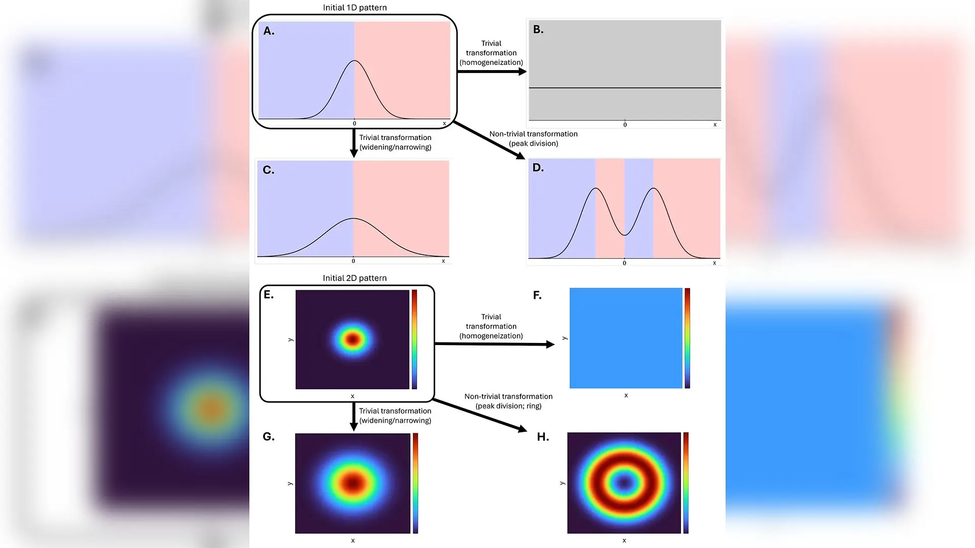
Pattern transformations. The figure shows examples of pattern transformations in 1D (A–D) and 2D (E–H). Panels represent different patterns (i.e. different spatial distributions of some gene product concentration g(t,x)\begin{document}$\boldsymbol{g}\left (t,\boldsymbol{x}\right)$\end{document}). (A) and (E) are the initial patterns being transformed into the resulting patterns in (B–D) and (F–H). The x-axis is cell position along a 1D array of cells and the y-axis is the concentration of a gene product. The transformation from (A) (resp., (E)) into (B) (resp., (F)) is trivial because the resulting pattern is homogeneous. The transformation from (A) (resp., (E)) to (C) (resp., (G)) is trivial because the concentration maximum is at the same spatial position in both patterns. The transformation from (A) (resp., (E)) to (D) (resp., (H)) is non-trivial because the resulting pattern is heterogeneous and the pattern in (D) has maxima that were not present in (A). The blue, gray, and pink fi Kevin Martinez Anhom et al. / eLife

A single fertilized egg manages to organize itself into an intricate organism through a fundamental biological puzzle, translating microscopic genetic instructions into a functional anatomy of tissues and organs. How cells with identical DNA end up in precise physical locations to form complex spatial patterns has long fascinated developmental biologists. Researchers have now demonstrated that despite the vast theoretical number of possible molecular wiring diagrams, nature is constrained to just three fundamental classes of gene networks capable of driving this spatial organization.

To decode these architectural rules, scientists combined rigorous mathematical proofs with reaction-diffusion models. The findings, published in eLife, reveal that pattern formation through extracellular signaling relies strictly on specific topological categories. The authors, Kevin Martinez Anhom and Isaac Salazar-Ciudad from the Centre de Recerca Matemàtica in Barcelona, analyzed how intracellular gene products interact with diffusible signals, known as morphogens or growth factors, which travel between neighboring cells.

The mathematical framework shows that any biological network capable of generating a non-trivial spatial pattern must incorporate positive regulatory loops combined with inhibitory interactions. Without positive feedback loops, gene concentrations would simply decay over time due to constant degradation by cellular proteases. Conversely, without negative interactions, tissues would remain entirely uniform and homogeneous.

By classifying these feedback motifs based on how cells respond to extracellular signals, the researchers found that all functional networks fall into three distinct architectural families. The first family consists of hierarchical networks, which feature positive intracellular loops and rely on directional signaling cascades without extracellular feedback loops. The second group comprises over-Turing networks, characterized by negative extracellular loops coupled with positive intracellular loops. The third category contains classical Turing networks, which rely on interactions between positive and negative extracellular loops.
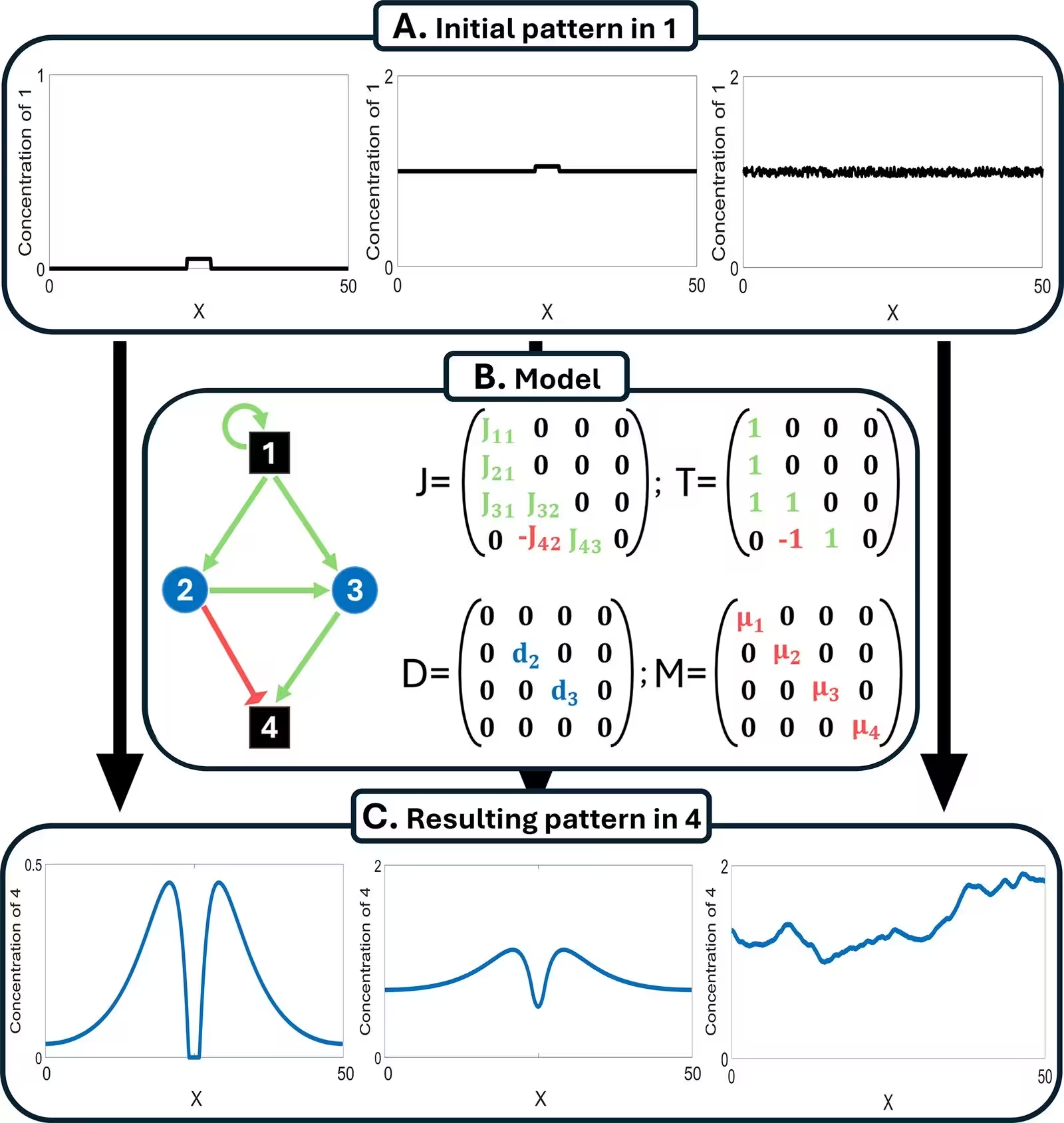
Figure 2: The model transforms initial patterns (A) into resulting patterns (C) through a set of equations implementing gene networks with extracellular signaling (B). The article considers three initial patterns (A): spike initial pattern (left), combined spike-homogeneous initial patterns (middle), and homogeneous-with-noise initial patterns, with small white noise (right). (B) Diagram of an example gene network. Black squares represent intracellular gene products, blue circles extracellular signals. Green arrows stand for positive regulations, while red arrows for negative regulations. Weights of the network are given by the J\begin{document}$\boldsymbol{J}$\end{document} matrix, while its topology by the T\begin{document}$\boldsymbol{T}$\end{document} matrix. D\begin{document}$\boldsymbol{D}$\end{document} represents the diffusion coefficients, and M\begin{document}$\boldsymbol{M}$\end{document} the degradation rates (see Equation 1). (C) Example resulting patterns for each initial pattern i
The model transforms initial patterns (A) into resulting patterns (C) through a set of equations implementing gene networks with extracellular signaling (B). The article considers three initial patterns (A): spike initial pattern (left), combined spike-homogeneous initial patterns (middle), and homogeneous-with-noise initial patterns, with small white noise (right). (B) Diagram of an example gene network. Black squares represent intracellular gene products, blue circles extracellular signals. Green arrows stand for positive regulations, while red arrows for negative regulations. Weights of the network are given by the J\begin{document}$\boldsymbol{J}$\end{document} matrix, while its topology by the T\begin{document}$\boldsymbol{T}$\end{document} matrix. D\begin{document}$\boldsymbol{D}$\end{document} represents the diffusion coefficients, and M\begin{document}$\boldsymbol{M}$\end{document} the degradation rates (see Equation 1). (C) Example resulting patterns for each initial pattern i Credit: Kevin Martinez Anhom et al. / eLife

The study highlights how each topological class dictates specific physical outcomes across one-dimensional and two-dimensional cellular arrays. Hierarchical networks produce radially symmetric patterns featuring highly customizable peaks and valleys, allowing for independent tuning of individual tissue structures. In contrast, Turing and over-Turing networks generate periodic waves or noisy configurations where parameters affect the entire pattern globally rather than locally.

These theoretical insights significantly narrow down the genetic mechanisms that experimental biologists must consider when studying embryonic development and organogenesis. By establishing that developmental gene networks can only evolve within a strictly limited set of architectural classes, the work provides a powerful predictive framework for understanding both natural anatomy and the evolutionary diversification of multicellular life.

The research was published in eLife on November 13, 2026.

Fact Checked

This article has been fact checked for accuracy, with information verified against reputable sources. Learn more about us and our editorial process.

Last reviewed on .

Article history

  • Latest version

Reference(s)

  1. Martinez Anhom, Kevin., et al. “The zoo of the gene networks capable of pattern formation by extracellular signaling.” eLife, vol. 14, November 13, 2026 eLife Sciences Publications, Ltd, doi: 10.7554/eLife.107563. <https://doi.org/10.7554/eLife.107563>.

Cite this page:

Sharma, Priya. “Mathematical Proof Reveals All Living Tissue Patterns Rely on Just Three Gene Network Families.” BioScience. BioScience ISSN 2521-5760, 15 November 2026. <https://www.bioscience.com.pk/en/subject/biology/the-zoo-of-the-gene-networks-capable-of-pattern-formation-by-extracellular-signaling>. Sharma, P. (2026, November 15). “Mathematical Proof Reveals All Living Tissue Patterns Rely on Just Three Gene Network Families.” BioScience. ISSN 2521-5760. Retrieved November 15, 2026 from https://www.bioscience.com.pk/en/subject/biology/the-zoo-of-the-gene-networks-capable-of-pattern-formation-by-extracellular-signaling Sharma, Priya. “Mathematical Proof Reveals All Living Tissue Patterns Rely on Just Three Gene Network Families.” BioScience. ISSN 2521-5760. https://www.bioscience.com.pk/en/subject/biology/the-zoo-of-the-gene-networks-capable-of-pattern-formation-by-extracellular-signaling (accessed November 15, 2026).
End of the article