Hidden Cone Photoreceptor Subtypes Discovered in Mouse and Primate Retinas
Advanced microscopy reveals double outer segments and rigid structural walls within mammalian color-sensing cells.
Mouse and baboon retinas harbor previously unknown subtypes of cone photoreceptors featuring double outer segments and distinct protein distribution patterns. Researchers mapping these cellular variations have uncovered unexpected structural complexity in the light-sensing cells responsible for color vision.
Vision relies on specialized light-detecting cells known as photoreceptors, divided into rods for dim light and cones for daylight and color. While studying adult mouse and baboon retinas using advanced immunomarkers and confocal microscopy, a research team identified previously unrecognized morphological and immunological subtypes of cones, including rare double cones in mice that possess twin outer segments. The findings were published in PLOS One, shedding new light on retinal architecture.
Uncovering Hidden Cone Varieties in Mammalian Eyes
Cone photoreceptors are typically classified by the specific light-absorbing photopigments they contain, such as long-wavelength, medium-wavelength, or short-wavelength opsins. In the study, investigators examined retinal slices and flat-mounts from 24 mice and 5 baboons to analyze cellular structures using specialized antibodies and a binding protein called peanut agglutinin, which labels the extracellular matrix sheath surrounding cone outer segments. In standard mouse cones, opsin proteins concentrate primarily within the outer segment, which is the light-catching tip of the cell. However, the team identified a subset representing about 7.6 percent of long- and medium-wavelength opsin-expressing cones and 5.7 percent of short-wavelength opsin-expressing cones that displayed elevated opsin levels throughout their somas and axons.
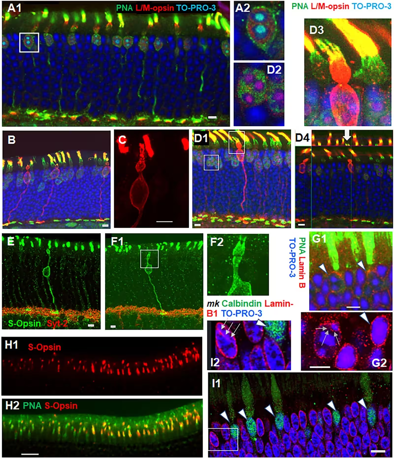
The researchers designated these specialized cells as lm-cones and ss-cones, respectively. Unlike regular photoreceptors, lm-cones feature significantly shorter and narrower outer segments along with rounder inner segments. Meanwhile, ss-cones display a long connecting stalk, known as a myoid, placing their cell bodies deeper within the outer nuclear layer among rod cell bodies. Furthermore, some of these specialized cones exhibited dual outer segments, pointing to the existence of mammalian double cones, a structural arrangement common in fish, reptiles, and birds but rarely documented in mammals.
Mapping S-Opsin Distribution and Structural Sheaths
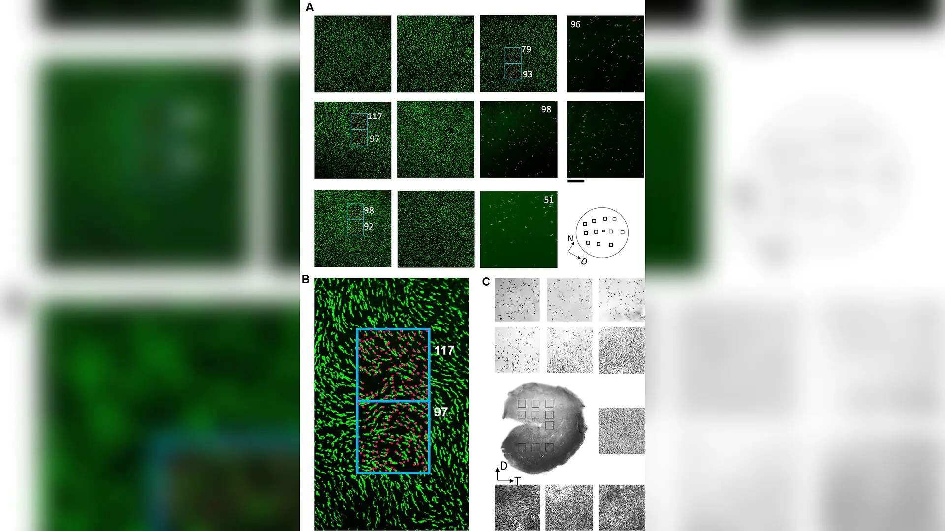
The study also mapped the precise spatial distribution of short-wavelength opsin cones across dark-adapted mouse retinas. By manually counting more than 150,000 cells per retina across 57 regions, the authors demonstrated that cone density follows a distinct Gaussian distribution. The highest concentrations appeared in the ventral-temporal region, located between 100 and 700 micrometers away from the optic nerve. Statistical modeling revealed that the dorsal-ventral density gradient was roughly twice as steep as the temporal-nasal gradient.
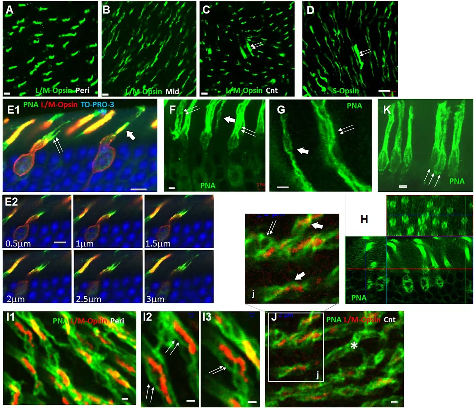
When examining the structural matrix surrounding primate and mouse cones, high-magnification imaging revealed intricate webs of parallel peanut agglutinin-positive strings and grids. In mouse retinas, outer segments featured either two intertwined strings or three to four thicker strands measuring roughly 0.25 to 0.35 micrometers in thickness. In baboon retinas, similar extracellular sheaths formed multi-barrel structures comprising up to 12 parallel walls that extended from the outer segment down to the inner segment. The researchers propose that these rigid protein sheaths act as mechanical walls, keeping delicate photoreceptors straight and structurally stable within the fluid environment of the eye.
The investigation further identified rare photoreceptor-retinal ganglion cell hybrids in mouse retinas, which combined a traditional outer segment with large cell bodies and long axons reaching deep into the inner plexiform layer. In baboon retinas, approximately 10.5 percent of cone outer segments expressed calcium-binding protein 5 instead of binding with peanut agglutinin, highlighting functional diversity across different retinal zones. While these observations provide a detailed anatomical baseline, the authors note that further functional research is necessary to understand how these newly described cone subtypes contribute to visual processing and whether their structural variations influence vulnerability to retinal degenerative diseases.
The research was published in PloS one on January 1, 2026.
This article has been fact checked for accuracy, with information verified against reputable sources. Learn more about us and our editorial process.
Last reviewed on .
Article history
- Latest version
Reference(s)
- Pang, Vivian Y.., et al. “The immunological subtypes of cone photoreceptors in mouse and primate retinas.” PLOS One, vol. 21, no. 11, November 13, 2026, pp. e0362261 Public Library of Science (PLoS), doi: 10.1371/journal.pone.0362261. <https://doi.org/10.1371/journal.pone.0362261>.
Cite this page:
- Posted by Hassan Raza