Targeting Brain Kinase Signaling Stops Severe Seizures in Mouse Tissue
Biology

Targeting Brain Kinase Signaling Stops Severe Seizures in Mouse Tissue

Blocking WNK-SPAK/OSR1 pathways forces neurons to clear out excess chloride ions, restoring inhibitory signaling and halting drug-resistant electrical storms.
By Hassan Raza
Published:
Email this Article
Targeting Brain Kinase Signaling Stops Severe Seizures in Mouse Tissue
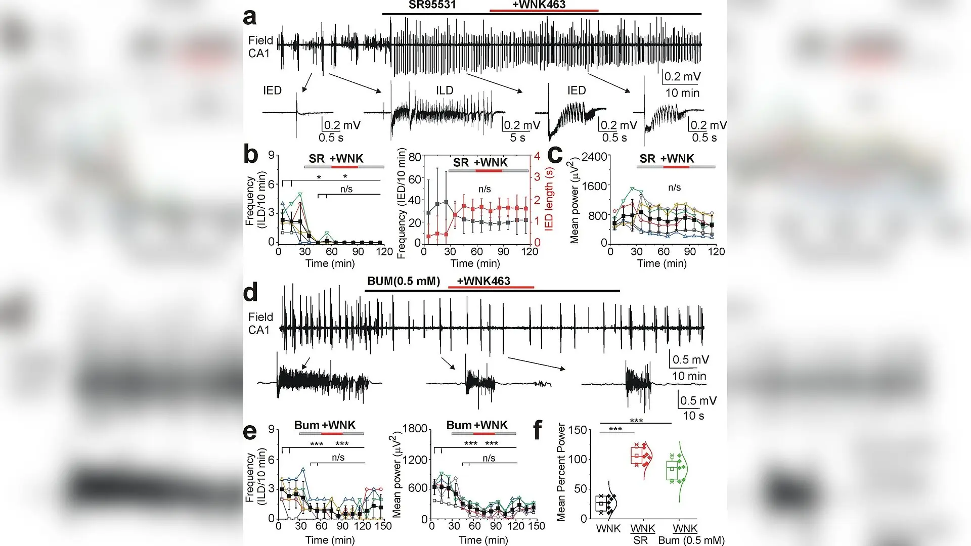
WNK463 had no effects in the presence of the GABAA-receptor antagonist and CCCs inhibitors. (a, d) Extracellular field potential recordings in the CA1 pyramidal cell layer in the organotypic hippocampal slices in vitro. Expansion of recurrent interictal (IEDs) and ictal (ILDs) epileptiform discharges in control and during drug applications. (b, c) Application of the GABAA-R antagonist SR95531 (10 µM) abolished spontaneous ILDs and induced large amplitude IEDs. Subsequent application of WNK463 (1 µM) in the presence of SR95531 did not change the mean frequency and duration of epileptiform discharges and corresponding power of electrical activity (N=6 slices; n/s – p>0.05; *p<0.05; Friedman RM ANOVA on Ranks, Dunn's test). (e) Cation-chloride cotransporter blocker bumetanide (0.5 mM) reduced the mean frequency of ILDs, corresponding power of electrical activity, and prevented the anti-ictal effects of WNK463 (N=6 slices; n/s – p>0.05; *p<0.05; ***p<0.001; one-way RM ANOVA, Tukey test). ( Volodymyr I Dzhala et al. / eLife

Mouse brain tissue experiments suggest that blocking a specific kinase signaling network can rapidly stop severe seizure activity by resetting how neurons manage chloride ions. Researchers observed that manipulating this pathway successfully eliminated recurrent seizure-like events in laboratory hippocampal slice cultures by forcing neurons to clear out excess salts.

When brain injuries or severe seizures occur, neurons often experience a dangerous influx of sodium and chloride ions. This ionic imbalance ruins the normal behavior of gamma-aminobutyric acid, which is the brain’s primary inhibitory neurotransmitter that normally keeps runaway electrical firing in check. Instead of calming neurons down, gamma-aminobutyric acid begins to excite them, fueling repetitive, prolonged seizures that resist standard medications.

How Brain Cells Manage Chloride Transport

Cellular chloride levels are strictly controlled by specialized molecular machines called cation-chloride cotransporters. One transporter, known as NKCC1, pumps chloride into cells, while another called KCC2 pumps it out. During acute brain trauma or continuous seizures, these transporters become heavily dysregulated through a signaling cascade controlled by WNK kinases and their downstream partners SPAK and OSR1. This kinase pathway phosphorylates the transporters, locking NKCC1 in an active state and KCC2 in an inactive state, which causes chloride to pile up inside neurons.
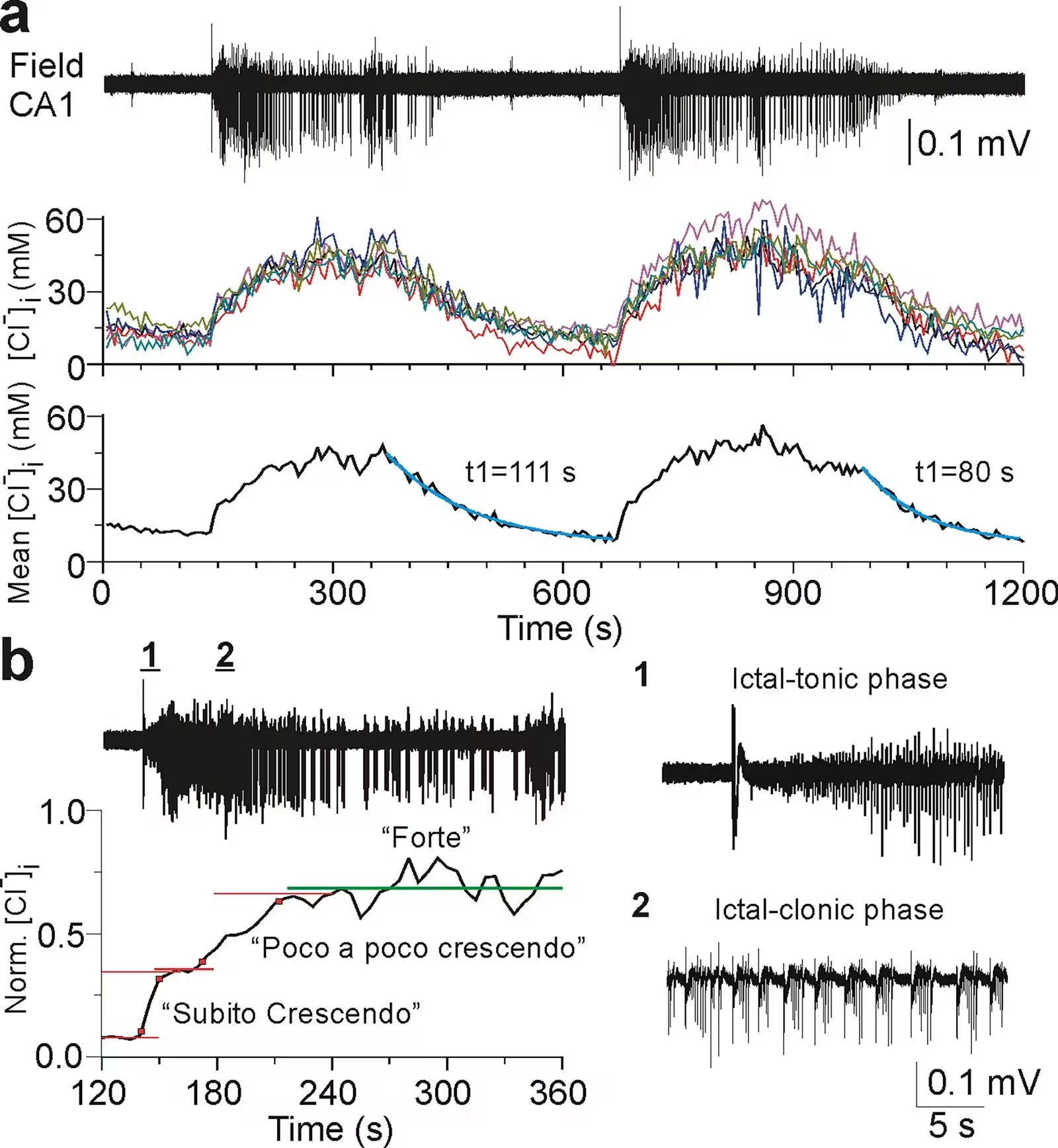
Figure 1: Neuronal chloride transients during spontaneous ictal-like discharges (ILDs). (a) Simultaneous extracellular field potential recording and two-photon fluorescence chloride imaging in the organotypic hippocampal slice at DIV18. Example of spontaneous recurrent ictal-like epileptiform discharges (ILDs) and corresponding [Cl−]i transients in the CA1 pyramidal cells (n=6), and the mean [Cl−]i. [Cl−]i progressively increased during ‘crescendo’ phase of sustained ictal-tonic discharges, was relatively stable during ‘forte’ phase of intermittent ictal-clonic discharges, and progressively decreased during ‘decrescendo’ phase of secondary after-discharges and post-ictal depression (decay time constant (t1) 80–111 s). (b) Expansion of ILDs and mean [Cl−]i changes normalized to values between 0 and 1 ([0, 1]). [Cl−]i rapidly increased during hyper-synchronous onset of ILD (‘subito crescendo’) and subsequent sustained ictal-tonic discharges (‘poco a poco crescendo’) and was relatively stable durin
Neuronal chloride transients during spontaneous ictal-like discharges (ILDs). (a) Simultaneous extracellular field potential recording and two-photon fluorescence chloride imaging in the organotypic hippocampal slice at DIV18. Example of spontaneous recurrent ictal-like epileptiform discharges (ILDs) and corresponding [Cl−]i transients in the CA1 pyramidal cells (n=6), and the mean [Cl−]i. [Cl−]i progressively increased during ‘crescendo’ phase of sustained ictal-tonic discharges, was relatively stable during ‘forte’ phase of intermittent ictal-clonic discharges, and progressively decreased during ‘decrescendo’ phase of secondary after-discharges and post-ictal depression (decay time constant (t1) 80–111 s). (b) Expansion of ILDs and mean [Cl−]i changes normalized to values between 0 and 1 ([0, 1]). [Cl−]i rapidly increased during hyper-synchronous onset of ILD (‘subito crescendo’) and subsequent sustained ictal-tonic discharges (‘poco a poco crescendo’) and was relatively stable durin Credit: Volodymyr I Dzhala et al. / eLife

To test whether reversing this process could halt seizures, a team of neuroscientists from Harvard Medical School and Massachusetts General Hospital examined the effects of a pharmacological compound called WNK463. This drug acts as an allosteric inhibitor of the WNK-SPAK/OSR1 signaling pathway. The findings were published in eLife, detailing how the treatment influenced neuronal chloride dynamics and electrical activity in mouse brain tissue.

Clearing Out Accumulated Ions to Stop Seizures

Using advanced two-photon microscopy combined with genetically encoded fluorescent chloride indicators, the researchers tracked real-time changes in intracellular chloride concentration within CA1 pyramidal neurons. Under control conditions, spontaneous ictal-like discharges triggered massive spikes in internal chloride. However, when the tissue was treated with 1 micromolar of WNK463, the drug progressively reduced interictal chloride levels, enhanced the rate of chloride extrusion, and shortened the duration of seizure episodes before eventually abolishing them altogether.

The experimental team discovered that WNK463 did not alter baseline chloride levels when all synaptic activity was blocked by tetrodotoxin, a neurotoxin that stops voltage-gated sodium channels. This distinction indicates that the inhibitor specifically targets activity-dependent chloride accumulation that happens during seizures rather than disrupting normal resting states. By simultaneously inhibiting NKCC1 influx and stimulating KCC2-mediated efflux, the treatment restored functional inhibition across the neural network.
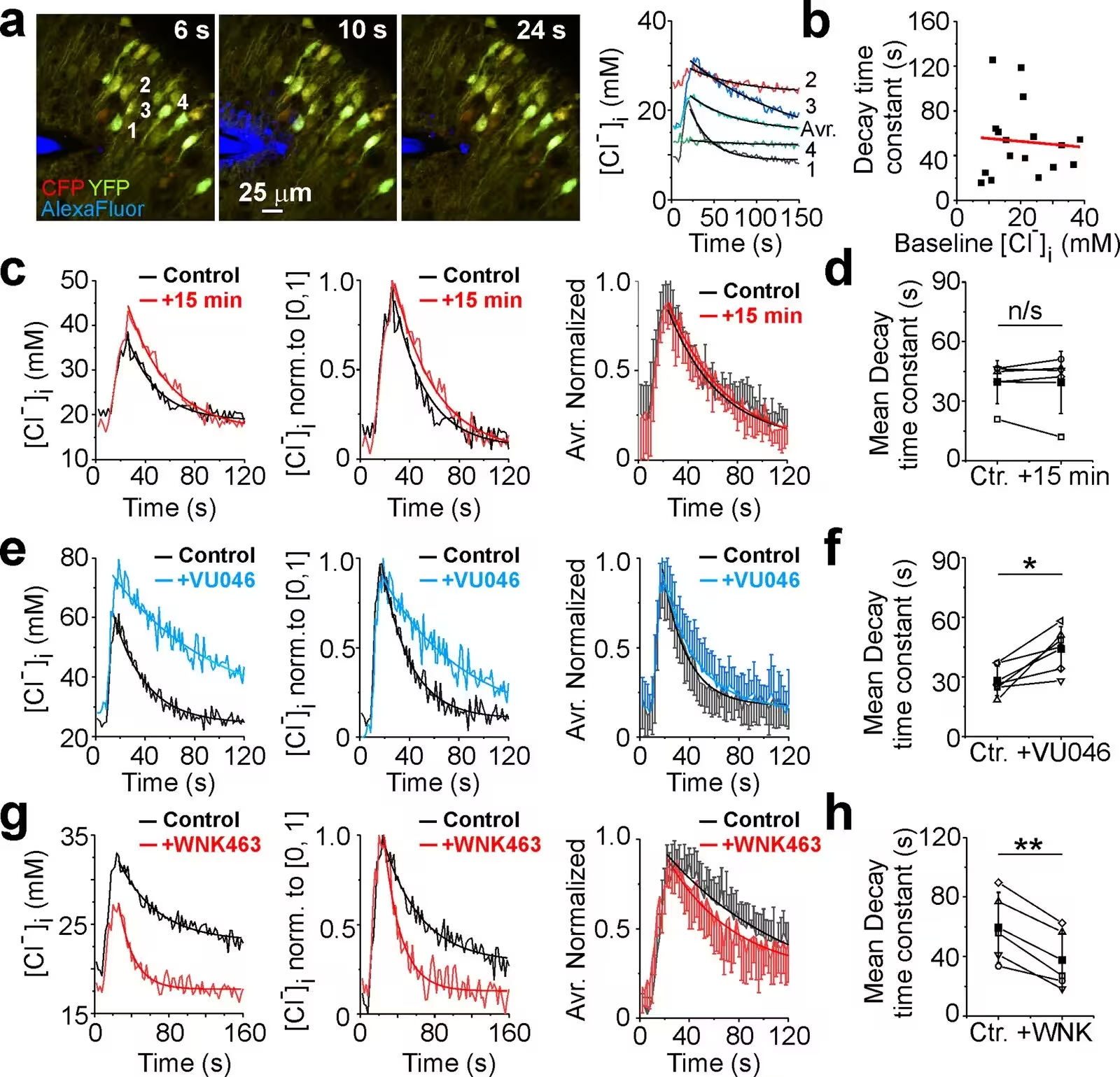
Figure 2: WNK-SPAK/OSR1 inhibitor WNK463 facilitated neuronal chloride extrusion. (a) An illustration of the designed experiment to induce neuronal chloride transients in the CA1 pyramidal cells expressing sCLM. The puff micropipette filled with extracellular solution containing GABA (50 µM) and L-Glu (50 µM) was placed above the dendritic area of the neurons. Merged CFP (red) and YFP (green) fluorescent signals in subpopulation of neurons before (6 s), during (10 s), and after (24 s) GABA/L-Glu puff application. Alexa Fluo-594 (blue) was added for visualization. Corresponding [Cl–]i transients induced by GABA/L-Glu puff application. Exponential decay fit (y=y0+A1exp(-(x-x0)/t1)) was used to measure decay time constant (t1) of [Cl-]i recovery in individual cells (1-4; solid curves). (b) Linear regression analysis revealed that decay time constant of [Cl−]i recovery in individual cells was independent of the baseline [Cl−]i and corresponding EGABA. (c, e, g) [Cl–]i transients, corresponding [Cl−]
WNK-SPAK/OSR1 inhibitor WNK463 facilitated neuronal chloride extrusion. (a) An illustration of the designed experiment to induce neuronal chloride transients in the CA1 pyramidal cells expressing sCLM. The puff micropipette filled with extracellular solution containing GABA (50 µM) and L-Glu (50 µM) was placed above the dendritic area of the neurons. Merged CFP (red) and YFP (green) fluorescent signals in subpopulation of neurons before (6 s), during (10 s), and after (24 s) GABA/L-Glu puff application. Alexa Fluo-594 (blue) was added for visualization. Corresponding [Cl–]i transients induced by GABA/L-Glu puff application. Exponential decay fit (y=y0+A1exp(-(x-x0)/t1)) was used to measure decay time constant (t1) of [Cl-]i recovery in individual cells (1-4; solid curves). (b) Linear regression analysis revealed that decay time constant of [Cl−]i recovery in individual cells was independent of the baseline [Cl−]i and corresponding EGABA. (c, e, g) [Cl–]i transients, corresponding [Cl−] Credit: Volodymyr I Dzhala et al. / eLife

Investigating Alternative Mechanisms and Limitations

To confirm whether the anti-seizure effects relied strictly on these two known transporters, the researchers performed additional tests using selective pharmacological blockers and small interfering RNA gene silencing to knock down Slc12a2 and Slc12a5 expression. Interestingly, blocking either NKCC1 or KCC2 individually did not prevent WNK463 from stopping the seizure-like discharges. The treatment only lost its full efficacy when both cotransporters were simultaneously inhibited, pointing toward cooperative interactions or the involvement of additional unidentified ion exchangers.

While these laboratory observations offer a promising therapeutic avenue for drug-resistant epilepsy and traumatic brain injury, the authors caution that organotypic slice cultures do not fully replicate the complex vascular supply and extracellular potassium regulation found in living brains. Future studies will need to evaluate whether WNK kinase inhibitors can safely and effectively control seizures in whole-animal models.

The research was published in eLife on November 17, 2026.

Fact Checked

This article has been fact checked for accuracy, with information verified against reputable sources. Learn more about us and our editorial process.

Last reviewed on .

Article history

  • Latest version

Reference(s)

  1. Dzhala, Volodymyr I., et al. “WNK-SPAK/OSR1 signaling pathway facilitates ictal activity via reduced neuronal chloride extrusion rate.” eLife, vol. 15, November 17, 2026 eLife Sciences Publications, Ltd, doi: 10.7554/eLife.112181. <https://doi.org/10.7554/eLife.112181>.

Cite this page:

Raza, Hassan. “Targeting Brain Kinase Signaling Stops Severe Seizures in Mouse Tissue.” BioScience. BioScience ISSN 2521-5760, 19 November 2026. <https://www.bioscience.com.pk/en/subject/biology/wnk-spak-osr1-signaling-pathway-facilitates-ictal-activity-via-reduced-neuronal-chloride-extrusion-rate>. Raza, H. (2026, November 19). “Targeting Brain Kinase Signaling Stops Severe Seizures in Mouse Tissue.” BioScience. ISSN 2521-5760. Retrieved November 19, 2026 from https://www.bioscience.com.pk/en/subject/biology/wnk-spak-osr1-signaling-pathway-facilitates-ictal-activity-via-reduced-neuronal-chloride-extrusion-rate Raza, Hassan. “Targeting Brain Kinase Signaling Stops Severe Seizures in Mouse Tissue.” BioScience. ISSN 2521-5760. https://www.bioscience.com.pk/en/subject/biology/wnk-spak-osr1-signaling-pathway-facilitates-ictal-activity-via-reduced-neuronal-chloride-extrusion-rate (accessed November 19, 2026).
End of the article