New Cryo-EM Preprocessing Method Boosts Protein Resolution
By correcting radiation-induced signal decay on a per-image basis before 3D reconstruction, researchers achieved sharper molecular maps.
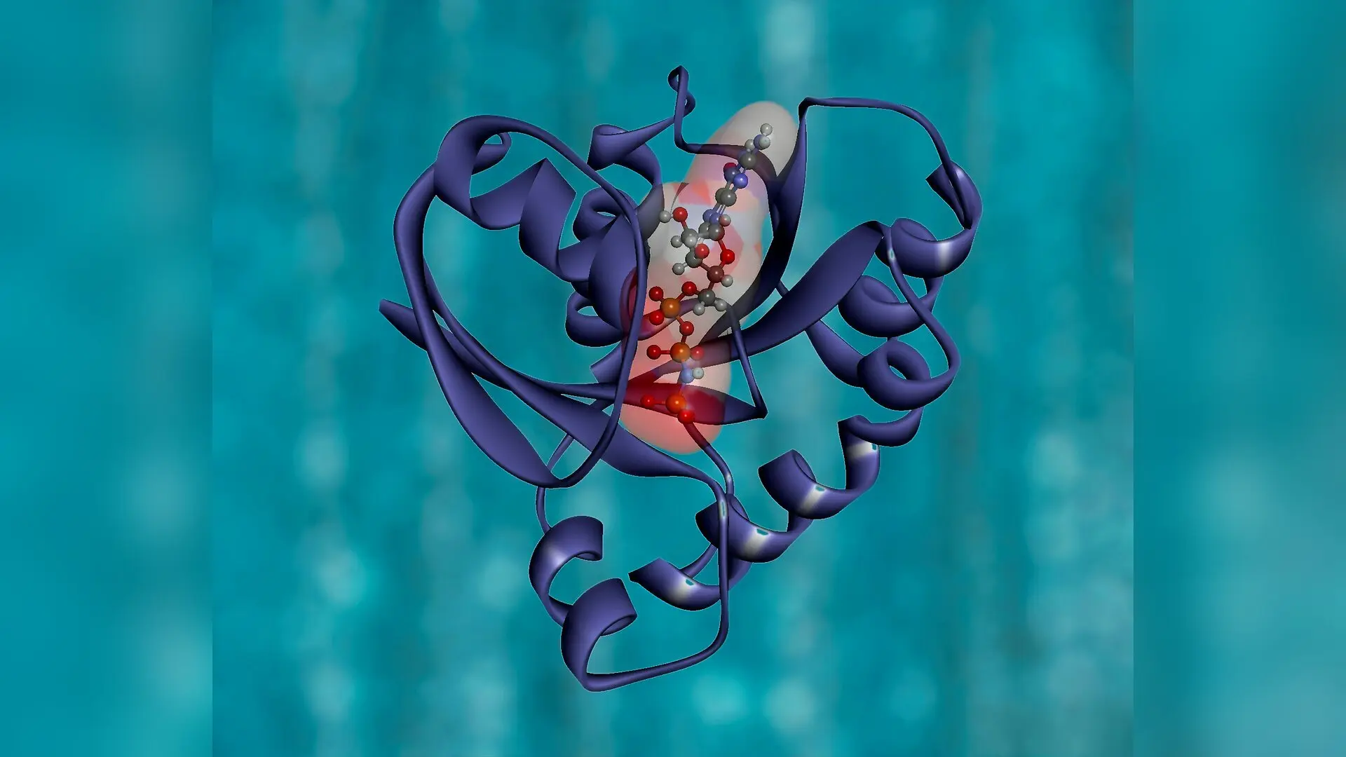
A new image processing technique for single-particle cryo-electron microscopy allows scientists to measure and correct radiation-induced signal decay before building three-dimensional models, yielding sharper molecular maps. By estimating sample degradation on a per-micrograph basis instead of applying a single blanket correction at the very end of data processing, researchers achieved noticeable improvements in final resolution across three major protein datasets.
Single-particle cryo-electron microscopy, commonly known as cryo-EM, has revolutionized structural biology by letting researchers image frozen biological macromolecules at near-atomic resolutions. In this technique, scientists flash high-energy electron beams through a thin layer of vitreous ice containing suspended proteins, capturing thousands of two-dimensional projections. These flat snapshots are then computationally combined to reconstruct the complex three-dimensional shapes of proteins, viruses, and cellular machinery.
However, the imaging process itself damages the delicate samples. High-energy electrons break chemical bonds, cause specimen motion, and introduce intense shot noise that blurs high-frequency details. This degradation causes an exponential decay in the amplitude and power spectra of protein images, mathematically modeled by a parameter called the B factor. Traditionally, laboratories tackle this signal loss during post-processing by flattening the power spectrum of the final 3D reconstruction map. This standard workflow assumes that every single experimental image in a dataset suffers the exact same degree of degradation.
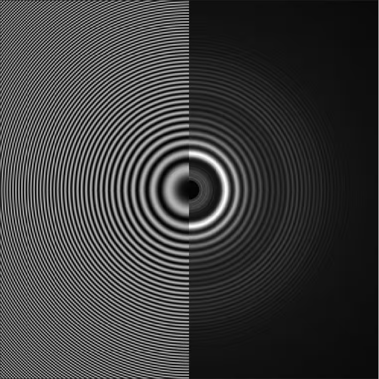
To overcome this limitation, applied mathematicians Ayelet Heimowitz and Amit Singer developed a preprocessing framework that calculates individual B factors directly for each micrograph. The details of the work are published in arXiv, outlining a method that isolates mid-high spatial frequencies where protein structures follow predictable statistical behavior known as Wilson statistics and Guinier law. By targeting specific frequencies where the microscope contrast transfer function reaches its maximum or minimum, the authors demonstrated that individual micrograph degradation can be precisely quantified prior to 3D modeling.
How Preprocessing Outperforms Traditional Post-Processing Workflows
The research team tested their modified pipeline on three publicly available real-world datasets from the Electron Microscopy Public Image Archive, including measurements of beta-galactosidase, the Plasmodium falciparum 80S ribosome, and the membrane protein arabinosyltransferase EmbB. For the beta-galactosidase sample containing 84 micrographs, calculating individual B factors with an average value of 95 square angstroms improved the final 3D reconstruction resolution from 4.4 angstroms down to 4.2 angstroms. Similar gains materialized for the parasite ribosome dataset, where accounting for unique micrograph degradation pushed resolution from 3.7 angstroms to 3.6 angstroms.
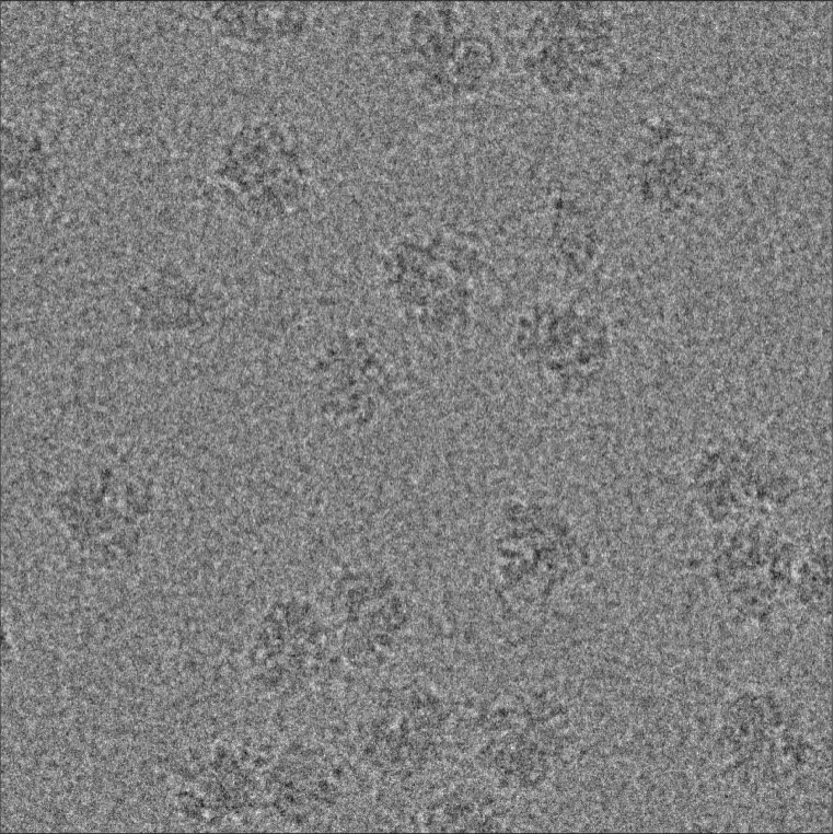
The most dramatic enhancement appeared in the arabinosyltransferase EmbB dataset, where correcting signal decay prior to classification enabled software to reliably spot and discard roughly 7,000 damaged projections. Removing these low-quality images catapulted the final structural resolution from 6.4 angstroms to 5.2 angstroms. The authors attribute the wide variability in B factors across individual micrographs to subtle mechanical shifts during data collection, noting that drum-like vibrations of the carbon support film affect motion correction quality differently from one snapshot to the next.
What This Means for Future Structural Biology Research
By moving B factor correction to the beginning of the computational pipeline, laboratories can clean up noisy data before sorting particles into classes. This early correction prevents damaged high-resolution signals from confusing classification algorithms, ensuring that only the highest quality data contributes to the final atomic model. While the approach requires additional computational steps before model building, the resulting boost in clarity could help scientists resolve elusive macromolecular structures that currently sit just out of reach.
The study was posted to the arXiv preprint server on November 18, 2026.
This article has been fact checked for accuracy, with information verified against reputable sources. Learn more about us and our editorial process.
Last reviewed on .
Article history
- Latest version
Reference(s)
- Heimowitz, Ayelet. “Micrograph B-factor estimation via Wilson statistics.” arXiv.org <http://arxiv.org/abs/2611.24150v1>.
Cite this page:
- Posted by Priya Sharma