Scientists Connected Three Human Brain Organoids And Witnessed A Primitive Form Of Learning
Researchers have successfully reshaped a three-organoid neural network using electrical stimulation, achieving clearer signal processing than ever before.
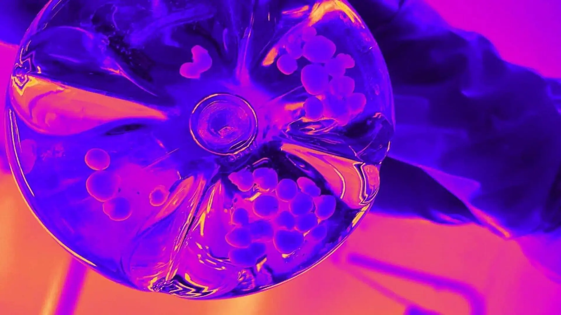
Researchers have successfully demonstrated that small clusters of human neural tissue, known as brain organoids, can undergo a form of functional reorganization when connected in specific modular configurations. By linking these miniature, three-dimensional neural networks and subjecting them to repeated electrical stimulation, scientists were able to trigger structural and functional changes that allowed the networks to better process and differentiate incoming signals.
Engineering Modular Neural Networks
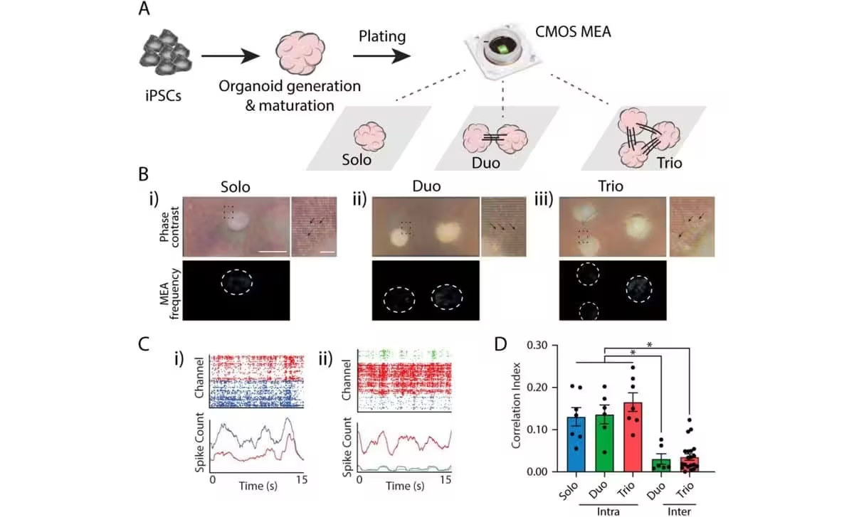
Brain organoids serve as vital models for neuroscientists, offering a way to study human neural processes that are otherwise inaccessible in living subjects. While these clusters of cells are not fully developed brains, they exhibit complex cellular organization and electrical signaling capabilities. To investigate how neural tissue evolves to handle specialized functions, a team led by neuroscientist Yoshiho Ikeuchi developed a platform using human induced pluripotent stem cells (hiPSCs).
The researchers cultivated these stem cells into neural tissue, which was then placed onto custom-designed chips equipped with microscopic electrodes. These electrodes served a dual purpose: they could both record electrical activity and deliver precise, targeted stimulation. The experimental architecture varied, with some chips hosting a single organoid, others two, and a subset featuring a trio of interconnected organoids. Over a period of two weeks, the organoids extended axons to forge connections with their neighbors, eventually establishing synchronized electrical communication across the multi-organoid modules.
The Threshold of Functional Learning
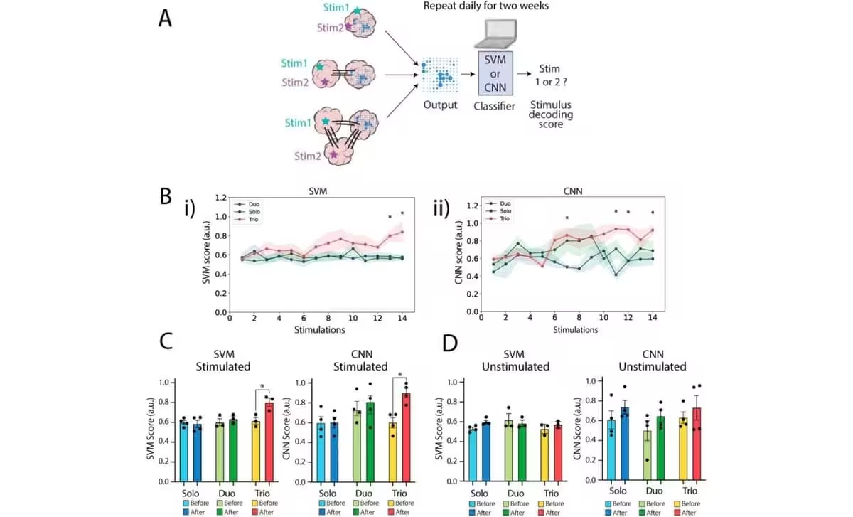
Following the development phase, the team initiated a two-week stimulation regimen. Daily, they applied electrical pulses at two distinct locations, recording the downstream activity to see if the network could distinguish between the input sources. As noted by ScienceAlert, the researchers intentionally chose stimulation sites that initially produced ambiguous, hard-to-differentiate responses.
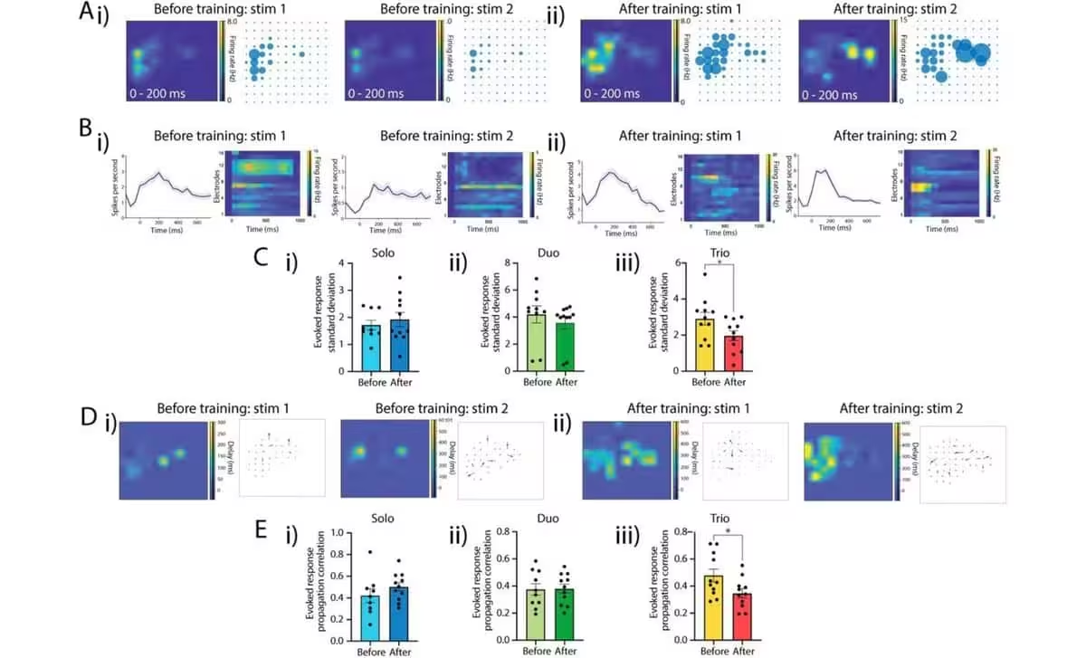
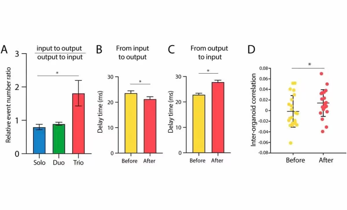
Using machine-learning algorithms to decode the neural output, the researchers discovered that the networks responded differently depending on their complexity. For single-organoid units and two-organoid pairs, the ability to distinguish between stimulation sites remained at chance levels even after the training period. However, the three-organoid networks showed a marked improvement. In these trios, the third, unstimulated organoid developed increasingly distinct, stable, and rapid neural responses that clearly correlated with which of the other two organoids received the initial signal. Control groups that were not exposed to the stimulation regimen showed no such improvement, confirming that the change was a result of active training rather than mere maturation.
Emergent Complexity in Biological Models
The findings suggest that a specific modular organization is required for neural tissue to adapt and perform specialized tasks. The researchers described the process as a rudimentary form of learning, where repeated input drives the rewiring of the network to create task-relevant differences in neural behavior.
“Together, these results demonstrate that repeated input to an organized modular network can rewire organoids generated under identical conditions into functionally differentiated modules,” the team noted in their findings, “thereby generating task-relevant heterogeneity that underlies consistent functional enhancement in vitro.”
While these organoids are not conscious entities or complete miniature brains, the experiment highlights a fundamental mechanism by which connected tissue gains sophisticated capabilities. Looking ahead, the researchers aim to explore how more complex signals might influence development. They also emphasize the importance of identifying potential “hub neurons” that may act as conductors, facilitating the communication necessary for networks to function as integrated systems. By integrating biochemical and molecular analyses with these electrical studies, the team hopes to further demystify the biological underpinnings of synaptic plasticity.
This article has been fact checked for accuracy, with information verified against reputable sources. Learn more about us and our editorial process.
Last reviewed on .
Article history
- Latest version
Reference(s)
- Starr, Michelle. “Scientists Connected 3 Human Mini-Brains in a Dish. They Learned Something The Others Couldn't..”, September 30, 2026 ScienceAlert <https://www.sciencealert.com/scientists-connected-3-human-mini-brains-in-a-dish-they-learned-something-the-others-couldnt>.
Cite this page:
- Posted by Hassan Raza