Bacterial Efflux Pump Gene Unveils Hidden Mutations in Drug-Resistant Pathogens
Analysis of clinical E. coli isolates reveals novel amino acid substitutions inside key transmembrane transport helices.
A common bacterial pump found in urinary tract infection pathogens carries surprising genetic variations that could change how drugs are handled inside the cell. Researchers analyzing ten multidrug-resistant Escherichia coli samples from patients in Iraq found that every single strain carries the emrD gene, a molecular transport system traditionally known for pushing toxic compounds out of bacterial cells.
The study, published in PLOS One, reveals unexpected genetic diversity in a system long considered a rigid housekeeping gene. While these bacterial strains showed full resistance to multiple antibiotic classes including penicillins, third-generation cephalosporins, and fluoroquinolones, the changes discovered within the pump’s structural framework highlight how these microscopic defenders might adapt.
How a 12-Helix Molecular Pump Operates
The emrD gene encodes a protein made of 12 transmembrane helices that form a central cavity inside the inner membrane of the bacteria. This channel uses energy from the cell’s electrochemical gradient to pump out harmful hydrophobic molecules and unwanted chemical agents. Past structural work established that the transporter acts like a rocker switch, tilting back and forth to move substrates from the interior to the outside environment.
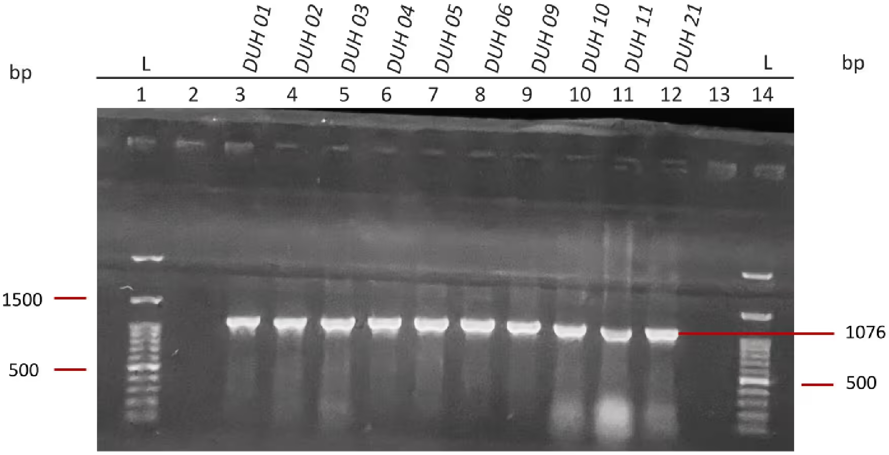
In this targeted pilot investigation, scientists extracted bacterial DNA from ten clinical samples collected at Azadi Teaching Hospital in Duhok. The team used polymerase chain reaction testing to successfully amplify the 1,076-base-pair emrD gene across all ten isolates, proving that the genetic blueprint for the efflux pump remains universally present even in highly resistant backgrounds.
What Revealed the Unexpected Evolutionary Divergence
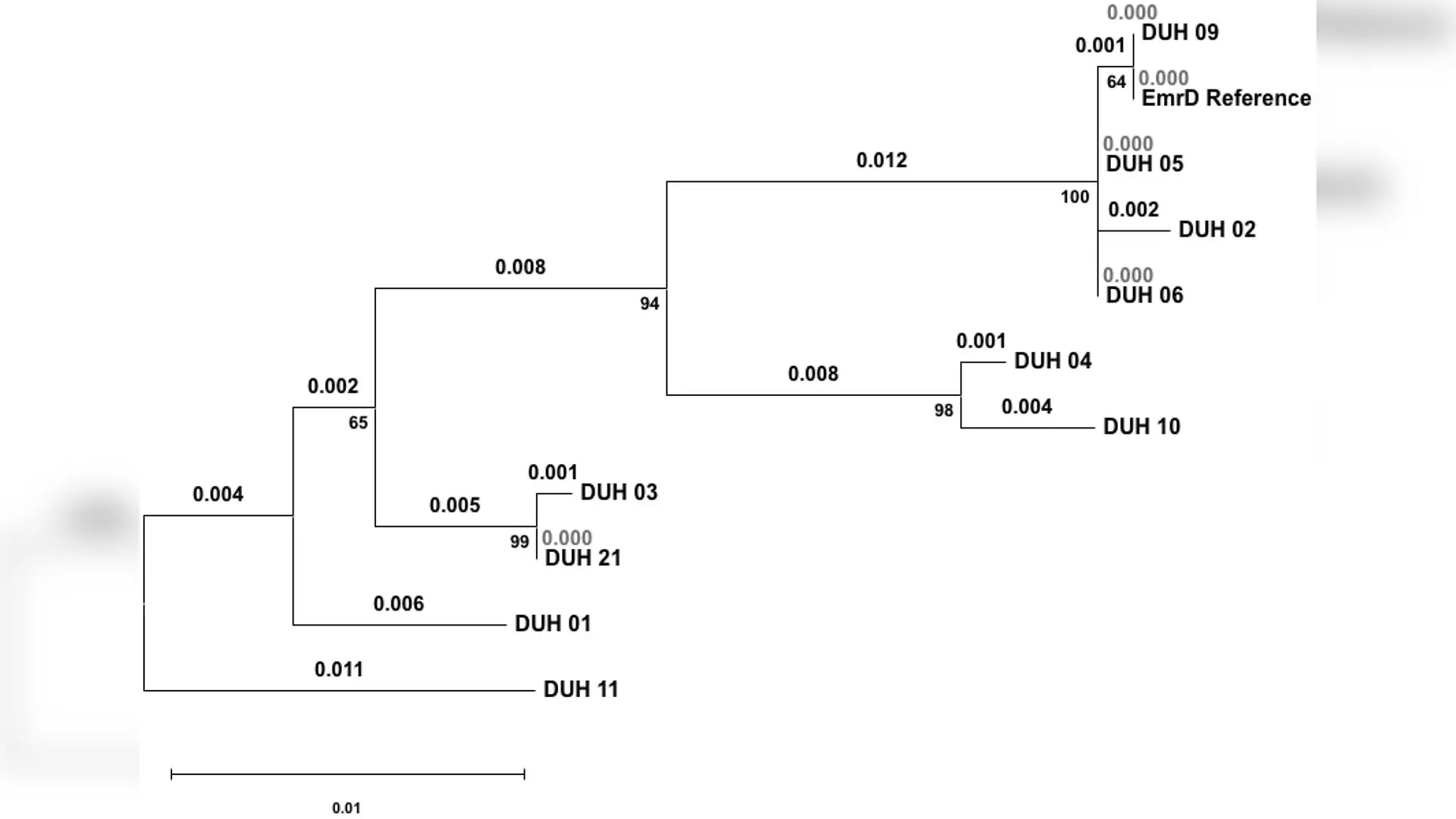
After sequencing the genetic material and translating the codons into protein sequences, the authors mapped the mutations against standard reference strains like K-12 MG1655 and uropathogenic reference lines CFT073 and 536. A phylogenetic tree constructed using the Hasegawa-Kishino-Yano substitution model divided the ten clinical strains into three distinct clusters alongside one highly divergent basal outlier designated as strain DUH 11.
Most strains grouped into Cluster I alongside standard reference sequences, but other samples showed noticeable separation. Strain DUH 11 stood out with the longest branch length in the dataset, indicating a significantly different genetic trajectory compared to the other local clinical isolates.
Which Specific Amino Acid Changes Altered the Transmembrane Helices
Protein translation uncovered three specific non-synonymous amino acid substitutions located directly within the core transport machinery. Strain DUH 10 displayed a valine-to-alanine change at position 170 within the sixth transmembrane helix, alongside a valine-to-isoleucine substitution at position 304 in the twelfth helix. Cross-strain comparisons confirmed that the latter substitution matches patterns found in established uropathogenic reference genomes.
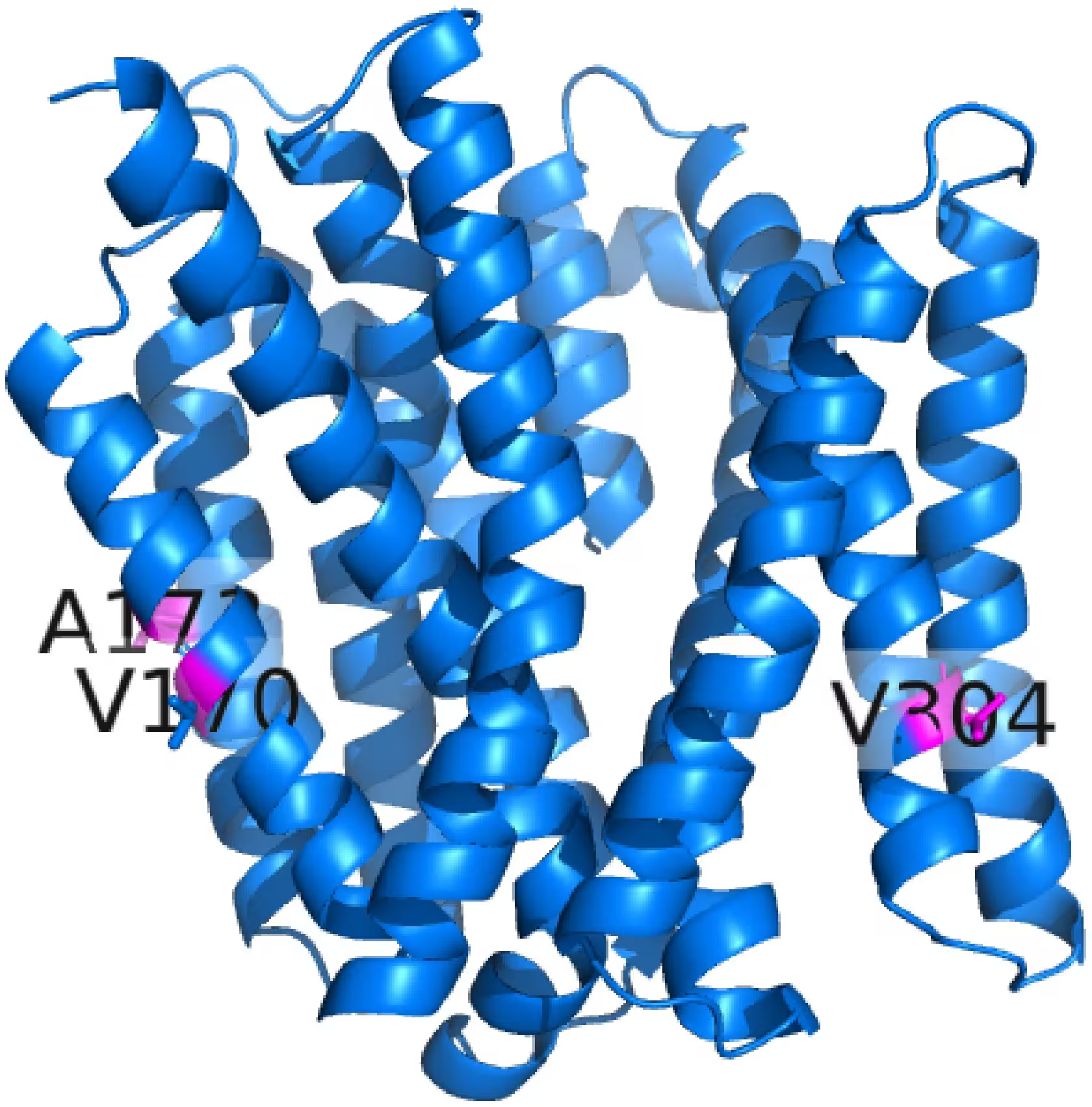
More strikingly, strain DUH 02 harbored an alanine-to-threonine substitution at position 173 inside the sixth transmembrane helix. This specific alteration introduces a polar residue in place of a non-polar one, representing a novel variant that did not appear in standard commensal strains or wider genomic databases. Because helices six and twelve line the main substrate translocation pathway, such localized shifts could alter the internal cavity volume and flexibility required for binding various drugs.
What These Resistance Profiles Mean for Future Treatments
All ten tested isolates also harbored blaCTX-M-1 and blaTEM genes associated with extended-spectrum beta-lactamase production, while 60 percent carried blaSHV. The simultaneous presence of these enzymatic defenses and structural efflux variations points toward cooperative survival strategies against complex drug regimens.
The authors note several limitations, including the small pilot sample size of ten isolates and the lack of molecular dynamics simulations or functional assays to measure exact minimum inhibitory concentrations for the novel mutant. Future research will need to test whether the A173T variant directly alters drug transport kinetics or cellular survival under antibiotic pressure.
The research was published in PLOS One on November 11, 2026.
This article has been fact checked for accuracy, with information verified against reputable sources. Learn more about us and our editorial process.
Last reviewed on .
Article history
- Latest version
Reference(s)
- Mahmood, Svala H.., et al. “Molecular diversity and structural profiling of the EmrD efflux pump in multidrug-resistant uropathogenic Escherichia coli.” PLOS One, vol. 21, no. 11, November 11, 2026, pp. e0362259 Public Library of Science (PLoS), doi: 10.1371/journal.pone.0362259. <https://doi.org/10.1371/journal.pone.0362259>.
Cite this page:
- Posted by Hassan Raza