Anemia Drug Triggers Colon Cancer Cell Death by Hitting an Unexpected Enzyme
Biology

Anemia Drug Triggers Colon Cancer Cell Death by Hitting an Unexpected Enzyme

Researchers find that Molidustat bypasses its intended target to exploit a metabolic vulnerability in mutated cells.
By Priya Sharma
Published:
Email this Article
Anemia Drug Triggers Colon Cancer Cell Death by Hitting an Unexpected Enzyme
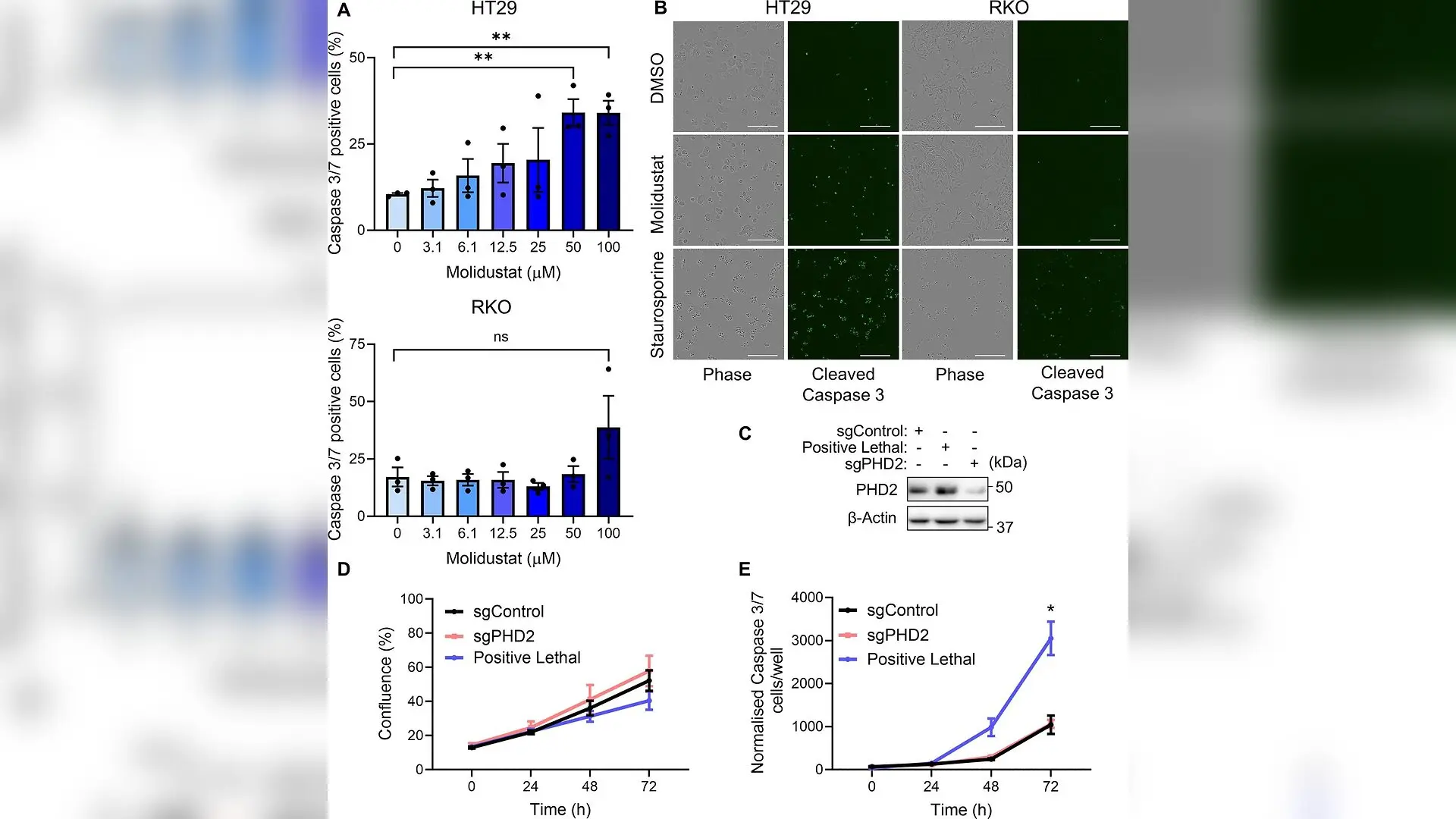
Molidustat induces cell death in colonic cell lines independently of PHD inhibition. (A) Percentage of cleaved Caspase-3/7 positive HT29 (top) and RKO (bottom) cells. Cells were treated with Molidustat for 48 hr at indicated concentrations; 10 μM Staurosporine was used as a positive control (100% cell death). Mean + SEM is assessed using unpaired two-tailed Student’s t-test, **p<0.01, (ns) non-significant. (B) Representative images of cleaved Caspase-3/7 signal in DMSO, Molidustat (90 μM), and Staurosporine-treated cells. Scale bar: 300 μm. (C) Representative western blot of PHD2 levels in HT29 cells. (D) Percentage confluency of HT29 cells post-transfection with the indicated guide RNAs. (E) Cleaved Caspase-3/7 signal in HT29 cells post-transfection with the indicated crRNAs. Mean + SEM is assessed using two-way ANOVA, *p<0.05. N=3 biologically independent experiments. Figure 1—source data 1.PDF file containing the original, uncropped western blots for Figure 1C, with the relevant ban Chiara Asselborn et al. / eLife

An anemia drug currently used in clinical settings triggers cell death in colon cancer cells by unexpectedly binding to a vital cellular detoxification enzyme rather than its intended target, according to a study published in eLife. Researchers discovered that Molidustat, a medication developed to inhibit prolyl hydroxylase domain proteins, selectively targets a metabolic vulnerability in cancer cells with mutations in the adenomatous polyposis coli gene.

Uncovering an Unexpected Cancer Drug Target

Mutations in the adenomatous polyposis coli gene appear in the vast majority of colorectal cancer cases, initiating uncontrolled cell growth by hyperactivating specific signaling pathways. Scientists have long explored oxygen-sensing enzymes known as prolyl hydroxylases, particularly PHD2, as therapeutic checkpoints to disrupt these cancer pathways. However, when investigators treated human colon cancer cell lines carrying adenomatous polyposis coli mutations with Molidustat, the drug induced significant cell death that standard PHD2 inhibition could not explain. Genetic knockout experiments revealed that deleting the PHD2 gene alone did not reduce cell viability or trigger apoptosis, pointing toward an off-target mechanism.

To identify where the drug was actually binding, the research team employed thermal proteome profiling and chemical proteomics using specialized affinity matrices. These proteome-wide screens revealed glutathione S-transferase P1, an enzyme responsible for neutralizing toxic compounds and managing cellular stress, as a primary off-target of Molidustat. Biochemical assays subsequently confirmed that Molidustat directly blocks the enzymatic activity of glutathione S-transferase P1, inducing a sharp surge in reactive oxygen species within the treated cells.
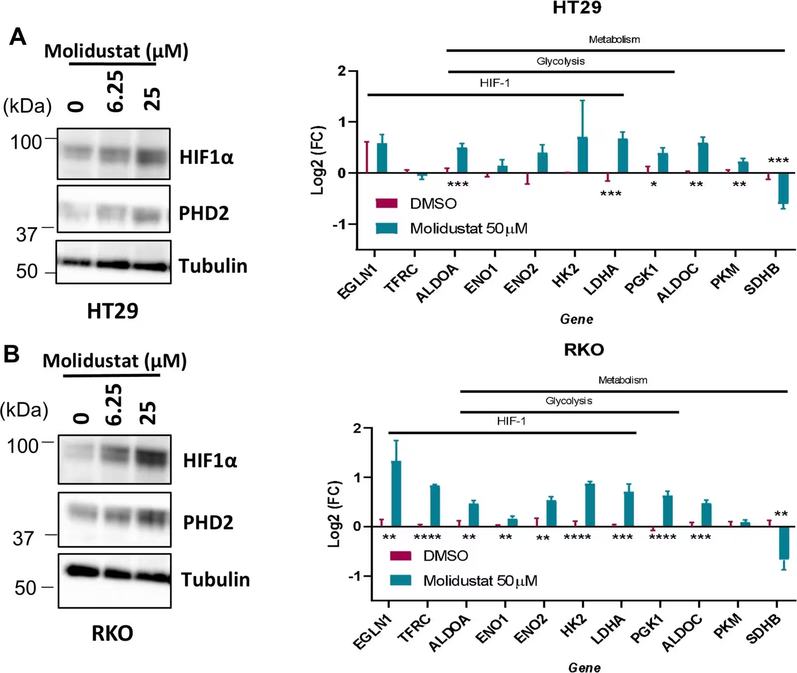
Figure 1—figure supplement 1: Molidustat induces HIF signalling in colonic cell lines. (A) HT29 cells treated with Molidustat for 16 hr. Left: Representative western blot of HIF1α and PHD2 at the indicated Molidustat concentrations (0, 6.25, 25 μM), with β-tubulin as a loading control. Right: Protein levels of HIF-responsive genes after treatment with 50 μM Molidustat, expressed as log2 fold change relative to DMSO. (B) As in A, for RKO cells. Mean + SEM is assessed using Student’s t-test, *p<0.05, **p<0.01, ***p<0.001, ****p<0.0001. N=3 biologically independent experiments. Figure 1—figure supplement 1—source data 1.PDF file containing the original, uncropped western blots for Figure 1—figure supplement 1A and B, with the relevant bands and molecular weight markers labelled. Figure 1—figure supplement 1—source data 2.Original image files for the western blot analyses displayed in Figure 1—figure supplement 1A and B.
Molidustat induces HIF signalling in colonic cell lines. (A) HT29 cells treated with Molidustat for 16 hr. Left: Representative western blot of HIF1α and PHD2 at the indicated Molidustat concentrations (0, 6.25, 25 μM), with β-tubulin as a loading control. Right: Protein levels of HIF-responsive genes after treatment with 50 μM Molidustat, expressed as log2 fold change relative to DMSO. (B) As in A, for RKO cells. Mean + SEM is assessed using Student’s t-test, *p<0.05, **p<0.01, ***p<0.001, ****p<0.0001. N=3 biologically independent experiments. Figure 1—figure supplement 1—source data 1.PDF file containing the original, uncropped western blots for Figure 1—figure supplement 1A and B, with the relevant bands and molecular weight markers labelled. Figure 1—figure supplement 1—source data 2.Original image files for the western blot analyses displayed in Figure 1—figure supplement 1A and B. Credit: Chiara Asselborn et al. / eLife

Cellular Collapse Driven by Combined Inhibition

To understand the downstream consequences of this dual engagement, the authors generated single and double CRISPR-Cas9 knockout models lacking PHD2, glutathione S-transferase P1, or both. Quantitative proteomics and metabolomics showed that simultaneous loss of both proteins produced profound metabolic and energetic imbalances. The double knockout cells suffered a high AMP-to-ATP ratio, severe shifts in cellular redox indicators such as the GSSG-to-GSH ratio, and widespread depletion of tricarboxylic acid cycle intermediates.

This metabolic crisis triggered a coordinated systems-level shift toward growth arrest and apoptotic cell death. Upregulated proteins included key markers of oxidative stress and mitochondrial apoptosis, such as mitochondrial effectors DIABLO and HTRA2, alongside the tumor suppressor PML. The proteomic changes closely mirrored the cellular damage observed when intact cancer cells were exposed directly to Molidustat.
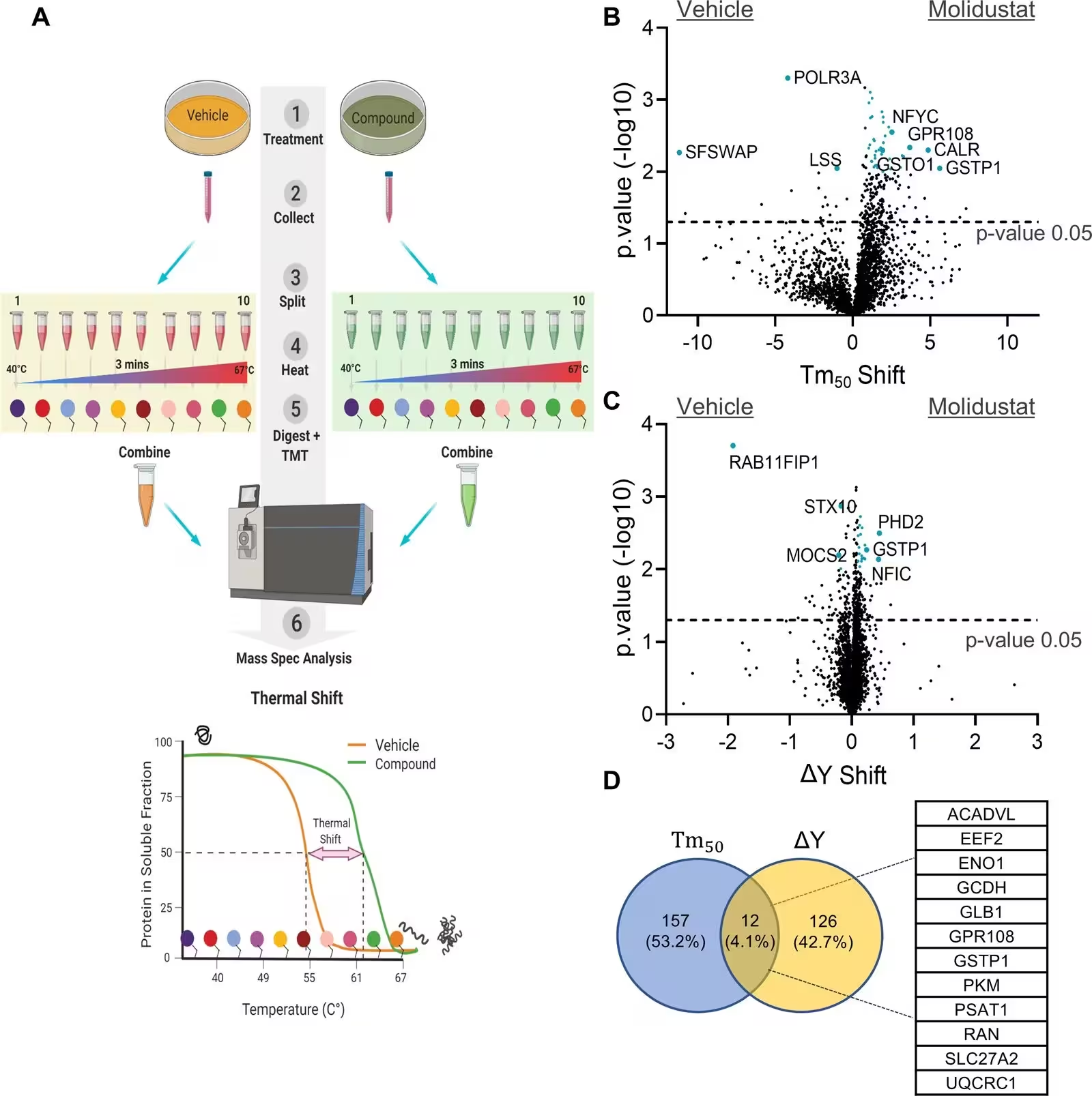
Figure 2: Thermal proteome profiling reveals multiple Molidustat targets. (A) Intact cells are treated with compound or vehicle control (1). Cells are collected (2), split into fractions (3) with each fraction heated to a different temperature (4). After heat treatment, cells are lysed and proteins are digested to peptides (5). The peptides in each fraction are labelled with a unique TMT tag, which can be analysed using mass spectrometer (6), generating melting curves and Tm50 values for the whole proteome. (B) Volcano plot of hits identified using the Tm50 method. Tm50 shift and –log10 p-value are shown. Proteins exhibiting a significant shift (<0.05) greater than 1°C were considered potential targets. (C) Volcano plot of all hits identified using the ΔY method. ΔY shift and –log10 p-value are shown. Proteins with a significant increase (<0.05) in ΔY greater than 0.1 were taken forward as potential hits. (D) Venn diagram showing the overlap of targets identified by both methods. Twelve proteins
Thermal proteome profiling reveals multiple Molidustat targets. (A) Intact cells are treated with compound or vehicle control (1). Cells are collected (2), split into fractions (3) with each fraction heated to a different temperature (4). After heat treatment, cells are lysed and proteins are digested to peptides (5). The peptides in each fraction are labelled with a unique TMT tag, which can be analysed using mass spectrometer (6), generating melting curves and Tm50 values for the whole proteome. (B) Volcano plot of hits identified using the Tm50 method. Tm50 shift and –log10 p-value are shown. Proteins exhibiting a significant shift (<0.05) greater than 1°C were considered potential targets. (C) Volcano plot of all hits identified using the ΔY method. ΔY shift and –log10 p-value are shown. Proteins with a significant increase (<0.05) in ΔY greater than 0.1 were taken forward as potential hits. (D) Venn diagram showing the overlap of targets identified by both methods. Twelve proteins Credit: Chiara Asselborn et al. / eLife

Testing Drug Sensitivity in Organoid Models

Because whole animal models faced drug delivery hurdles in reaching the colon effectively, the team evaluated Molidustat using murine intestinal organoids derived from genetically engineered mouse models. These stem-cell-derived structures faithfully reproduce the epithelial organization of human tissues. Organoids harboring adenomatous polyposis coli mutations or KRAS alterations displayed marked, dose-dependent reductions in viability when treated with Molidustat, accompanied by a sharp rise in caspase activity.

By contrast, wild-type control organoids and cultures treated with other structurally distinct hydroxylase inhibitors did not experience the same selective vulnerability. The findings suggest that Molidustat exploits a synthetic lethal interaction between glutathione S-transferase P1 and PHD2. This interaction opens new avenues for designing targeted combination therapies that harness oxidative stress vulnerabilities in specific colorectal cancer subtypes.

The research was published in eLife on November 10, 2026.

Fact Checked

This article has been fact checked for accuracy, with information verified against reputable sources. Learn more about us and our editorial process.

Last reviewed on .

Article history

  • Latest version

Reference(s)

  1. Asselborn, Chiara., et al. “Molidustat targets a synthetic lethal vulnerability in APC-mutant colorectal cancer through GSTP1 and PHD2 co-inhibition.” eLife, vol. 15, November 10, 2026 eLife Sciences Publications, Ltd, doi: 10.7554/eLife.110991. <https://doi.org/10.7554/eLife.110991>.

Cite this page:

Sharma, Priya. “Anemia Drug Triggers Colon Cancer Cell Death by Hitting an Unexpected Enzyme.” BioScience. BioScience ISSN 2521-5760, 11 November 2026. <https://www.bioscience.com.pk/en/subject/biology/molidustat-targets-a-synthetic-lethal-vulnerability-in-apc-mutant-colorectal-cancer-through-gstp1-and-phd2-co-inhibition>. Sharma, P. (2026, November 11). “Anemia Drug Triggers Colon Cancer Cell Death by Hitting an Unexpected Enzyme.” BioScience. ISSN 2521-5760. Retrieved November 11, 2026 from https://www.bioscience.com.pk/en/subject/biology/molidustat-targets-a-synthetic-lethal-vulnerability-in-apc-mutant-colorectal-cancer-through-gstp1-and-phd2-co-inhibition Sharma, Priya. “Anemia Drug Triggers Colon Cancer Cell Death by Hitting an Unexpected Enzyme.” BioScience. ISSN 2521-5760. https://www.bioscience.com.pk/en/subject/biology/molidustat-targets-a-synthetic-lethal-vulnerability-in-apc-mutant-colorectal-cancer-through-gstp1-and-phd2-co-inhibition (accessed November 11, 2026).
End of the article