Scientists Reveal How Cell Division Hubs Are Policed by Protein Destruction
Biology

Scientists Reveal How Cell Division Hubs Are Policed by Protein Destruction

Researchers map a dual-part molecular signal that directs TRIM37 to prune centrosome scaffolding proteins and prevent lethal mitotic arrest.
By Priya Sharma
Published:
Email this Article
Scientists Reveal How Cell Division Hubs Are Policed by Protein Destruction
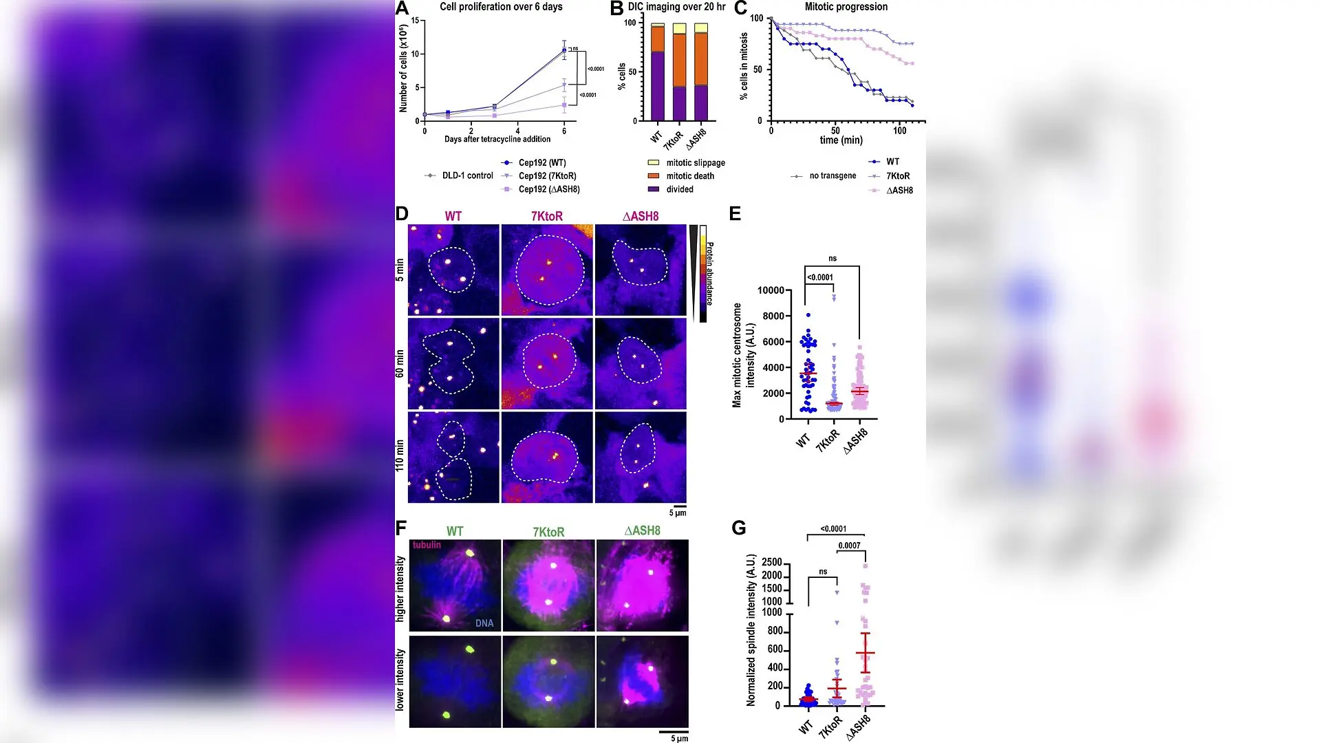
Cells expressing ubiquitination-deficient Cep192 mutants arrest in mitosis. (A) Quantification of cell proliferation during expression of Cep192 constructs (mean ± 95% CI; n = 6 biological replicates). P values (day 6 viability) from one-way ANOVA followed by post hoc Dunnett’s multiple comparison. (B) Quantification of cell fate after 20 h of live imaging (n ≥ 54 cells, three biological replicates). (C) Time-course quantification showing the cumulative percentage of cells completing mitosis over time with and without indicated Cep192 construct expression (n ≥ 20 cells, three biological replicates). (D) Representative images of cells expressing Cep192 constructs in mitosis over time. Cells are outlined in white. (E) Quantification of maximum fluorescence intensity of GFP-Cep192 constructs at mitotic centrosomes (median ± 95% CI; n ≥ 50 centrosomes, four biological replicates). P values from Kruskal–Wallis test, followed by post hoc Dunn’s multiple comparison. (F) Immunofluorescence of Weronika E Stachera et al. / The Journal of cell biology

Human cells rely on a precise molecular pruning system to keep their major microtubule-organizing centers at the right size, and researchers have finally uncovered the exact trigger that controls this process. Centrosomes act as the command hubs for cellular division, but when the scaffolding protein Cep192 builds up unchecked, it causes severe mitotic arrest and cell death. A team of cell biologists set out to decode how the enzyme TRIM37 targets Cep192 for destruction, discovering a dual-part signal that governs protein stability and guards against chromosomal instability.

The study, published in The Journal of Cell Biology, demonstrates that the ubiquitin ligase TRIM37 directly recognizes a specific sequence on the C terminus of the massive 307 kilodalton Cep192 protein. Lead author Weronika E. Stachera and colleagues established an in vitro ubiquitination assay using purified components from baculovirus-infected insect cells to prove direct interaction. Mass spectrometry analysis revealed that TRIM37 attaches molecular tags to a cluster of seven specific lysine residues located near the very end of Cep192, effectively marking the protein for proteasome-mediated degradation.

What is the bipartite degron that triggers destruction?

To pinpoint how the cellular machinery recognizes its target, the researchers mapped the docking interface on Cep192. They found that a composite region consisting of an intrinsically disordered region paired with a structured beta-barrel domain known as ASH8, collectively called IDR plus ASH8, is strictly required for stable binding. When the team mutated this recognition domain or altered the seven target lysines into arginines, Cep192 accumulated to abnormally high levels inside human colorectal cells, preventing normal protein turnover.
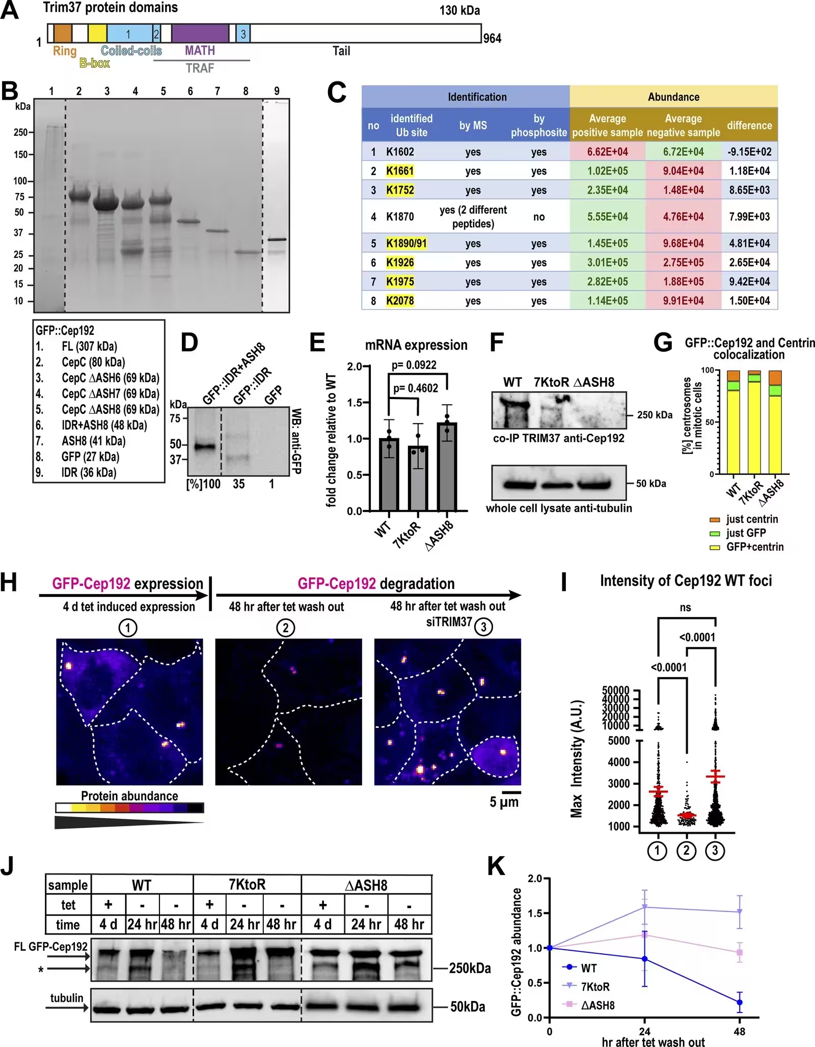
Figure S1: Identification of ubiquitinated residues and the TRIM37-binding site on Cep192. (A) Structured and conserved domains of TRIM37. (B) SDS-PAGE gel showing purified full-length Cep192 and fragments. (C) List of ubiquitinated lysines identified by MS and reported in PhosphoSitePlus (https://www.phosphosite.org/). (D) Western blot showing binding efficiency of Cep192 constructs to TRIM37. Constructs were purified from Escherichia coli and then pulled down using beads affixed to anti-TRIM37 antibody. Percentages represent relative binding capacity normalized to minimal TRIM37 binding region IDR+ASH8. (E) qPCR analysis of construct expression. Mean ± 95% CI; three biological replicates; P values from one-way ANOVA, followed by post hoc Dunnett’s multiple comparison. (F) Western blot showing binding efficiency of Cep192 constructs to TRIM37. Constructs were expressed in DLD-1 cells, followed by immunoprecipitation using beads coated with an anti-TRIM37 antibody. Tubulin from the total cell lys
Identification of ubiquitinated residues and the TRIM37-binding site on Cep192. (A) Structured and conserved domains of TRIM37. (B) SDS-PAGE gel showing purified full-length Cep192 and fragments. (C) List of ubiquitinated lysines identified by MS and reported in PhosphoSitePlus (https://www.phosphosite.org/). (D) Western blot showing binding efficiency of Cep192 constructs to TRIM37. Constructs were purified from Escherichia coli and then pulled down using beads affixed to anti-TRIM37 antibody. Percentages represent relative binding capacity normalized to minimal TRIM37 binding region IDR+ASH8. (E) qPCR analysis of construct expression. Mean ± 95% CI; three biological replicates; P values from one-way ANOVA, followed by post hoc Dunnett’s multiple comparison. (F) Western blot showing binding efficiency of Cep192 constructs to TRIM37. Constructs were expressed in DLD-1 cells, followed by immunoprecipitation using beads coated with an anti-TRIM37 antibody. Tubulin from the total cell lys Credit: Weronika E Stachera et al. / The Journal of cell biology

Further experiments showed that this specialized recognition module acts as a universal disposal ticket. When the authors fused the IDR plus ASH8 domain onto unrelated proteins such as the microtubule end-binding protein EB1 or monomeric green fluorescent protein, those proteins were rapidly degraded by TRIM37. This discovery confirms that the domain functions as a bona fide degron, capable of converting non-targeted proteins into substrates without requiring centrosome localization or higher-order protein clumping.

How do structural interactions drive high-affinity binding?

Biochemical assays and cross-linking mass spectrometry shed light on the physical architecture of the TRIM37 and Cep192 complex. The binding occurs with a strong mid-nanomolar affinity, showing an apparent dissociation constant of 75 nanomolar. Cross-linking analysis indicated that two separate coiled-coil domains on TRIM37 engage the compact beta-barrel structure of ASH8 and its preceding linker. This high-affinity interaction contrasts sharply with how TRIM37 handles other targets like Centrobin, which relies on low-affinity binding through its MATH domain to destroy large protein condensates.

When the degradation-resistant Cep192 mutants were overexpressed in human cells, the consequences were severe. Cell proliferation dropped by roughly 50 percent for the seven-lysine mutant and by about 75 percent for the domain deletion mutant. Live-cell imaging revealed that affected cells experienced prolonged metaphase arrest lasting hours instead of the normal 40-minute window, frequently culminating in mitotic failure or cell death.
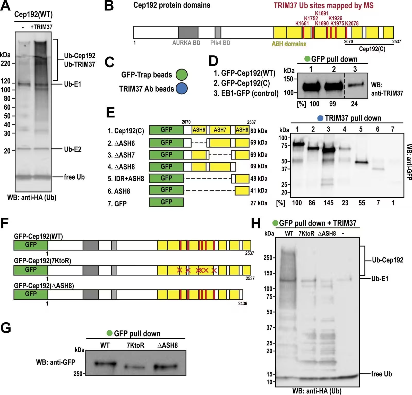
Figure 1: TRIM37 directly binds the IDR+ASH8 domain of Cep192 and ubiquitinates it on seven specific residues. (A) Western blot against HA-tagged ubiquitin from reconstituted ubiquitination reactions containing 10 µM HA-ubiquitin, 100 nM E1 UBE1, 100 nM E2 UbcH5b/UBE2D2, 1 µM Cep192, and either 0 µM (control) or 1 µM TRIM37. Representative of three replicates. (B) Schematic of Cep192 domains with ubiquitinated residues identified by MS. (C) Graphic representation of beads used for immunoprecipitation assays. Green: GFP-Trap magnetic particles; blue: beads conjugated to TRIM37 antibody. (D) Western blot showing TRIM37 binding to Cep192 constructs. Percentages represent relative binding capacity normalized to full-length Cep192. (E) Anti-GFP western blot showing Cep192 fragments that bind TRIM37. Percentages represent binding capacity normalized to the Cep192 C-terminal fragment. Representative of three replicates. (F) Cep192 constructs expressed in DLD-1 cells. (G) Western blot against GFP showin
TRIM37 directly binds the IDR+ASH8 domain of Cep192 and ubiquitinates it on seven specific residues. (A) Western blot against HA-tagged ubiquitin from reconstituted ubiquitination reactions containing 10 µM HA-ubiquitin, 100 nM E1 UBE1, 100 nM E2 UbcH5b/UBE2D2, 1 µM Cep192, and either 0 µM (control) or 1 µM TRIM37. Representative of three replicates. (B) Schematic of Cep192 domains with ubiquitinated residues identified by MS. (C) Graphic representation of beads used for immunoprecipitation assays. Green: GFP-Trap magnetic particles; blue: beads conjugated to TRIM37 antibody. (D) Western blot showing TRIM37 binding to Cep192 constructs. Percentages represent relative binding capacity normalized to full-length Cep192. (E) Anti-GFP western blot showing Cep192 fragments that bind TRIM37. Percentages represent binding capacity normalized to the Cep192 C-terminal fragment. Representative of three replicates. (F) Cep192 constructs expressed in DLD-1 cells. (G) Western blot against GFP showin Credit: Weronika E Stachera et al. / The Journal of cell biology

What are the broader implications for cancer and rare diseases?

The findings carry significant clinical relevance because dysregulation of TRIM37 is directly tied to human pathology. The genomic locus for TRIM37 is frequently amplified in several aggressive human malignancies, including neuroblastoma, breast cancer, liver tumors, and kidney cancer, where elevated enzyme levels promote chemoresistance. Conversely, loss-of-function mutations in the gene cause Mulibrey nanism, a rare autosomal recessive growth disorder characterized by severe prenatal and postnatal growth failure.

By mapping the exact structural rules that allow TRIM37 to recognize and degrade essential centrosome components, the research opens new avenues for understanding mitotic fidelity. Although the study utilized transgene overexpression models that require cautious interpretation, identifying this bipartite recognition motif suggests that similar mixed-architecture degrons may exist across the human proteome, waiting to be uncovered.

The research was published in The Journal of cell biology on January 4, 2027.

Fact Checked

This article has been fact checked for accuracy, with information verified against reputable sources. Learn more about us and our editorial process.

Last reviewed on .

Article history

  • Latest version

Reference(s)

  1. Stachera, Weronika E.., et al. “TRIM37 recognizes a bipartite degron to ubiquitinate centrosome substrates.” Journal of Cell Biology, vol. 226, no. 1, November 10, 2026 Rockefeller University Press, doi: 10.1083/jcb.202608031. <https://doi.org/10.1083/jcb.202608031>.

Cite this page:

Sharma, Priya. “Scientists Reveal How Cell Division Hubs Are Policed by Protein Destruction.” BioScience. BioScience ISSN 2521-5760, 11 November 2026. <https://www.bioscience.com.pk/en/subject/biology/trim37-recognizes-a-bipartite-degron-to-ubiquitinate-centrosome-substrates>. Sharma, P. (2026, November 11). “Scientists Reveal How Cell Division Hubs Are Policed by Protein Destruction.” BioScience. ISSN 2521-5760. Retrieved November 11, 2026 from https://www.bioscience.com.pk/en/subject/biology/trim37-recognizes-a-bipartite-degron-to-ubiquitinate-centrosome-substrates Sharma, Priya. “Scientists Reveal How Cell Division Hubs Are Policed by Protein Destruction.” BioScience. ISSN 2521-5760. https://www.bioscience.com.pk/en/subject/biology/trim37-recognizes-a-bipartite-degron-to-ubiquitinate-centrosome-substrates (accessed November 11, 2026).
End of the article