Laser Light Scattering Reveals Early Tumor Treatment Resistance
Label-free Raman spectroscopy tracks molecular shifts and metabolic rewiring during cancer therapy.
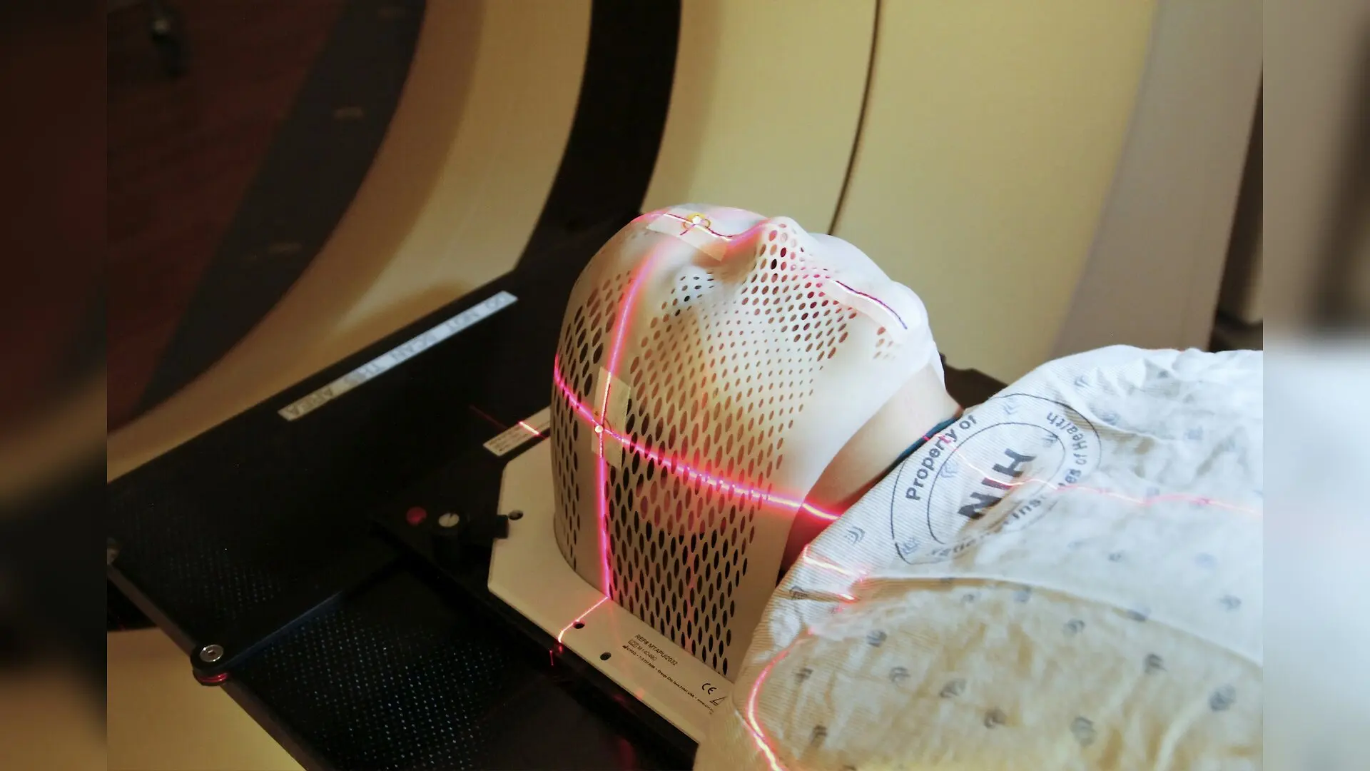
Using light scattering to peer inside living cells, scientists have tracked how aggressive tumors reorganize their internal chemistry to survive toxic cancer treatments. The work shows that measuring microscopic vibrations using laser light can reveal treatment resistance long before tumors actually shrink or change shape on conventional clinical scans.
Catching Molecular Shifts Before Tumors Change Shape
Monitoring how cancers respond to treatment remains one of modern oncology’s greatest hurdles. Standard clinical imaging methods like computed tomography scans or magnetic resonance imaging primarily assess tumor burden by measuring macroscopic changes in size and shape. However, critical molecular alterations associated with treatment sensitivity, drug resistance, or recurrence often arise much earlier, remaining undetected by traditional histopathology or imaging.
To overcome these limitations, researchers are turning to photonic techniques that probe intrinsic biochemical composition without adding dyes or labels. Raman spectroscopy, an analytical method based on the inelastic scattering of light by molecular vibrations, offers a label-free and non-destructive window into the biochemical makeup of cells, tissues, and biofluids. When a monochromatic laser beam interacts with a sample, a tiny fraction of the scattered photons undergoes an energy exchange corresponding to specific molecular bonds, producing a characteristic spectral fingerprint.
Recent advances in these technologies are detailed in a comprehensive study published in Chemical Society Reviews by Alcolea-Rodriguez and colleagues. The review synthesizes developments in Raman-based cancer therapy assessment from 2010 to 2026, bridging the gap between fundamental biophysics and translational oncology.
Decoding the Vibrational Signatures of Therapeutic Stress
The versatility of Raman scattering stems from its ability to capture holistic molecular profiles encompassing proteins, lipids, nucleic acids, and metabolites. Depending on the biological question, researchers can employ different modalities, ranging from conventional spontaneous Raman spectroscopy to high-speed coherent techniques like stimulated Raman scattering and coherent anti-Stokes Raman scattering.
When tumors are exposed to therapeutic interventions such as chemotherapy, radiotherapy, targeted drugs, or immunotherapy, they undergo profound biochemical rearrangements. Successful treatment is frequently marked by the breakdown of nucleic acids, visible as a decrease in the O-P-O backbone phosphodiester bands around 785 cm−1 and 1096 cm−1, alongside protein denaturation and membrane disruption.
Conversely, therapy-resistant cancer cells exhibit active metabolic and structural adaptation. For example, radiation-resistant or drug-tolerant cells often accumulate lipid droplets and alter their extracellular matrix composition, showing prominent changes in CH2 stretching vibrations near 2850 cm−1 and collagen-associated peaks. These shifts reflect metabolic rewiring and membrane remodeling that help cells endure therapeutic stress.
Navigating the Analytical Pipeline for Clinical Translation
Translating complex spectral data into clinically actionable insights requires standardized experimental design, robust preprocessing, and advanced multivariate data analysis. Because raw Raman spectra are susceptible to instrumental noise, background fluorescence, and substrate interference, standardized pipelines involving cosmic-ray removal, baseline correction, and intensity calibration are essential.
Dimensionality reduction techniques such as principal component analysis and non-negative matrix factorization help extract key biochemical features from hyperspectral datasets. When paired with supervised classification models like partial least squares discriminant analysis, support vector machines, or deep learning architectures, these spectral patterns can reliably differentiate between responsive and resistant tumor phenotypes.
Despite these technological strides, several hurdles remain on the path to routine clinical adoption. Inter-instrument variability, the need for standardized calibration protocols, and the validation of machine-learning models across multi-center cohorts require ongoing community-wide efforts such as open-access data repositories and standardized data reporting frameworks.
Moving Toward Precision Oncology and Real-Time Guidance
As Raman systems evolve from bulky laboratory setups into portable fiber-optic probes and ultrafast intraoperative imaging platforms, their clinical utility is expanding. Intraoperative guidance for surgical margin delineation represents one of the most mature applications, enabling real-time biochemical feedback during tumor resections. In parallel, liquid biopsy applications analyzing dried blood plasma or biofluids are emerging as promising tools for minimally invasive, longitudinal therapy monitoring.
Ultimately, integrating label-free Raman spectroscopy with advanced artificial intelligence and established clinical workflows could provide oncologists with a powerful complementary tool. By uncovering early treatment resistance and mapping tumor heterogeneity at the molecular level, these photonic approaches may help tailor therapeutic strategies and improve outcomes for cancer patients.
The research was published in Chemical Society reviews on November 16, 2026.
This article has been fact checked for accuracy, with information verified against reputable sources. Learn more about us and our editorial process.
Last reviewed on .
Article history
- Latest version
Reference(s)
- Alcolea-Rodriguez, Victor., et al. “Label-free Raman spectroscopy for cancer therapy response: from methodological framework to clinical translation.” Chemical Society Reviews, vol. 55, no. 22, January 1, 2026, pp. 12122-12189. Royal Society of Chemistry (RSC), doi: 10.1039/d6cs00966b. <https://doi.org/10.1039/d6cs00966b>.
Cite this page:
- Posted by Priya Sharma