Advanced 3D Tissue Models Transform Nanomedicine Preclinical Testing
New approach methodologies and organoids are replacing flat cell cultures to evaluate nanoparticle safety and drug delivery with greater human relevance.
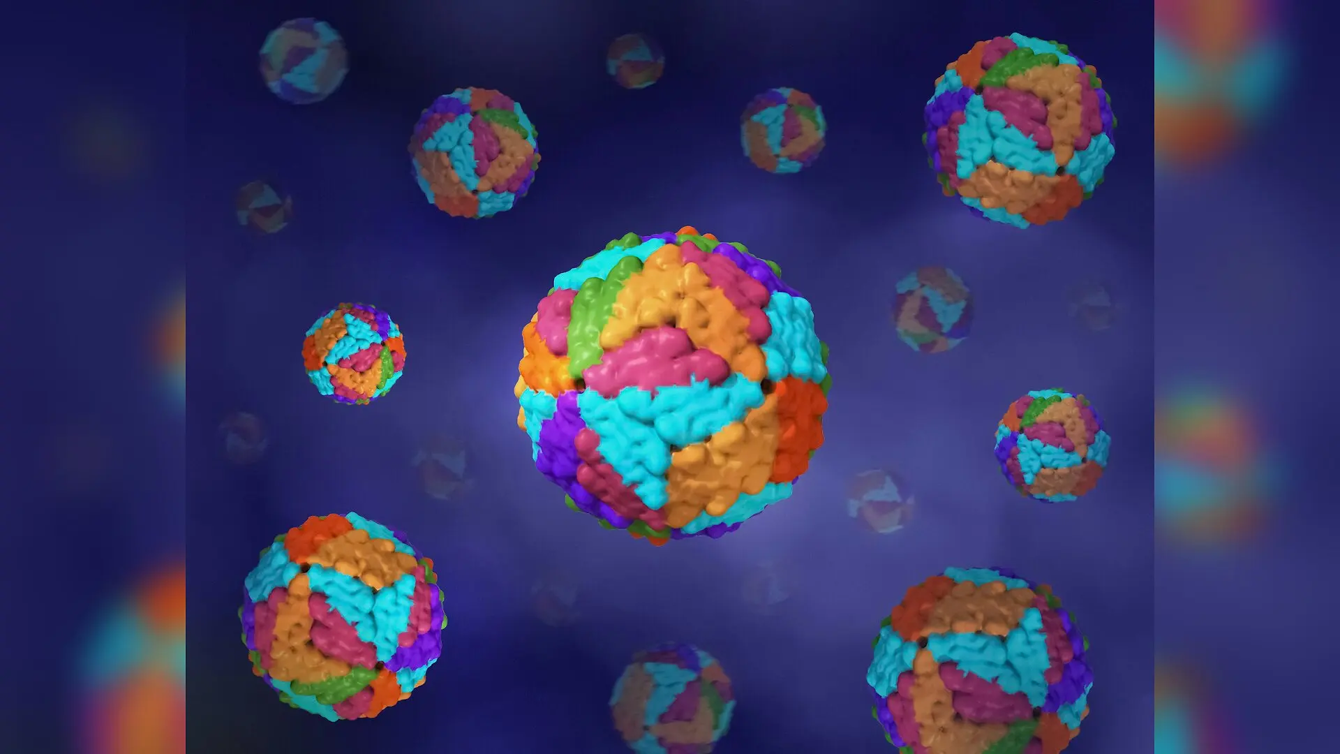
Tiny engineered structures less than one micrometer across are transforming modern medicine, yet testing their safety remains a major bottleneck for researchers. Over the past two decades, approximately 80 nanomedicine products have gained commercial approval from regulatory agencies, relying heavily on traditional flat laboratory cell cultures and animal testing during early screening. However, these conventional flat models fail to capture the complex architecture and fluid dynamics of living human tissues. To overcome these limitations, scientists are increasingly turning to three-dimensional tissue models and computational tools to evaluate how engineered particles behave inside the human body. These advanced testing platforms, collectively known as new approach methodologies, are detailed in a comprehensive evaluation published in Chemical Society Reviews by Yasmine Radwan and colleagues.
Why Flat Cell Cultures Fall Short in Nanomedicine
Conventional laboratory experiments typically grow cells in flat, two-dimensional monolayers that lack the structural complexity and surrounding extracellular matrix found in living organisms. In these flat setups, nutrients distribute evenly, and cells do not experience the mechanical forces or cell-to-cell contacts that dictate real biological responses. Consequently, observations made in flat cultures often fail to predict how nanoparticles will actually perform inside a living patient. This mismatch frequently necessitates expensive, time-consuming animal studies that raise ethical concerns and suffer from species-specific biological differences.
To bridge this gap, three-dimensional culture models such as multicellular spheroids, patient-derived organoids, and organ-on-a-chip microfluidic devices have emerged over the last decade. These systems allow cells to form natural three-dimensional architectures, deposit their own extracellular matrix, and establish nutrient and oxygen gradients similar to those seen in living tissues. By incorporating these physiologically relevant features, researchers can better investigate how nanoscale therapeutics penetrate dense barriers and interact with specific cell populations.
How Organic and Inorganic Nanoparticles Behave in Three Dimensions
Engineered nanoparticles fall into two broad categories, organic and inorganic, each presenting unique behaviors when tested in complex three-dimensional environments. Organic carriers include lipid-based nanoparticles, polymeric systems, and programmable nucleic acid nanoparticles assembled from RNA or DNA. In three-dimensional tumor models, polymeric and lipid carriers encounter diffusion limitations and dense extracellular matrices that slow their penetration, yielding uptake kinetics and therapeutic efficiencies that much more closely resemble actual clinical outcomes than flat cultures do.
Inorganic nanoparticles composed of gold, silver, iron oxide, silica, and quantum dots exhibit distinct accumulation patterns and clearance challenges across various organs. For instance, ultrasmall gold nanoparticles measuring less than two nanometers have demonstrated the ability to cross biological barriers like the blood-brain barrier in advanced spheroid models. Meanwhile, silver nanoparticles and quantum dots often show heightened toxicity in three-dimensional cultures compared to flat monolayers, largely because prolonged exposure times in structured tissues reveal degradation-dependent hazards that shorter assays miss.
Probing Organ-Specific Barriers and Toxicity Risks
Advanced tissue models have allowed scientists to investigate nanoparticle interactions across critical human organs, including the brain, liver, kidney, and placenta. In brain research, multicellular spheroids incorporating neurons, astrocytes, and endothelial cells have been used to track how surface chemistry dictates particle entry past protective barriers. Similarly, three-dimensional liver microtissues exposed to repeated low doses of metal oxide nanoparticles revealed cumulative functional impairments, such as declining albumin secretion, even when cell viability remained largely unaffected.
Kidney proximal tubule organoids have similarly improved the detection of nephrotoxicity caused by specific dendrimers and gold formulations by successfully reproducing key transport and biomarker responses. In placental models, microfluidic chips under continuous fluid flow demonstrated that shear stress and cellular differentiation significantly alter nanoparticle uptake and barrier integrity. These organ-specific findings indicate that functional and barrier endpoints often respond to stress long before overt cell death occurs, highlighting the superior sensitivity of three-dimensional systems.
Integrating Computational Models with Experimental Platforms
Pairing advanced laboratory models with computational techniques is becoming essential for managing the vast design space of nanoparticle formulations. Machine learning models, quantitative structure-activity relationships, and physiologically based pharmacokinetic simulations help prioritize candidate designs, predict immunological responses, and estimate delivered doses. By combining computational predictions with exposure-aware data from organ-on-a-chip and organoid platforms, researchers can streamline preclinical screening, reduce reliance on animal testing, and accelerate the clinical translation of precision nanomedicines.
The research was published in Chemical Society reviews on November 16, 2026.
This article has been fact checked for accuracy, with information verified against reputable sources. Learn more about us and our editorial process.
Last reviewed on .
Article history
- Latest version
Reference(s)
- Radwan, Yasmine., et al. “New approach methodologies in nanomedicine: bridging the gap between in silico , in vitro , and in vivo systems.” Chemical Society Reviews, vol. 55, no. 22, January 1, 2026, pp. 11707-11738. Royal Society of Chemistry (RSC), doi: 10.1039/d6cs01323f. <https://doi.org/10.1039/d6cs01323f>.
Cite this page:
- Posted by Hassan Raza