What Is MCH in a Blood Test? High, Low & Normal Ranges
Learn what MCH measures, the normal range, causes of high and low MCH, and how it helps diagnose iron deficiency, B12 deficiency, thalassemia, and other blood disorders.
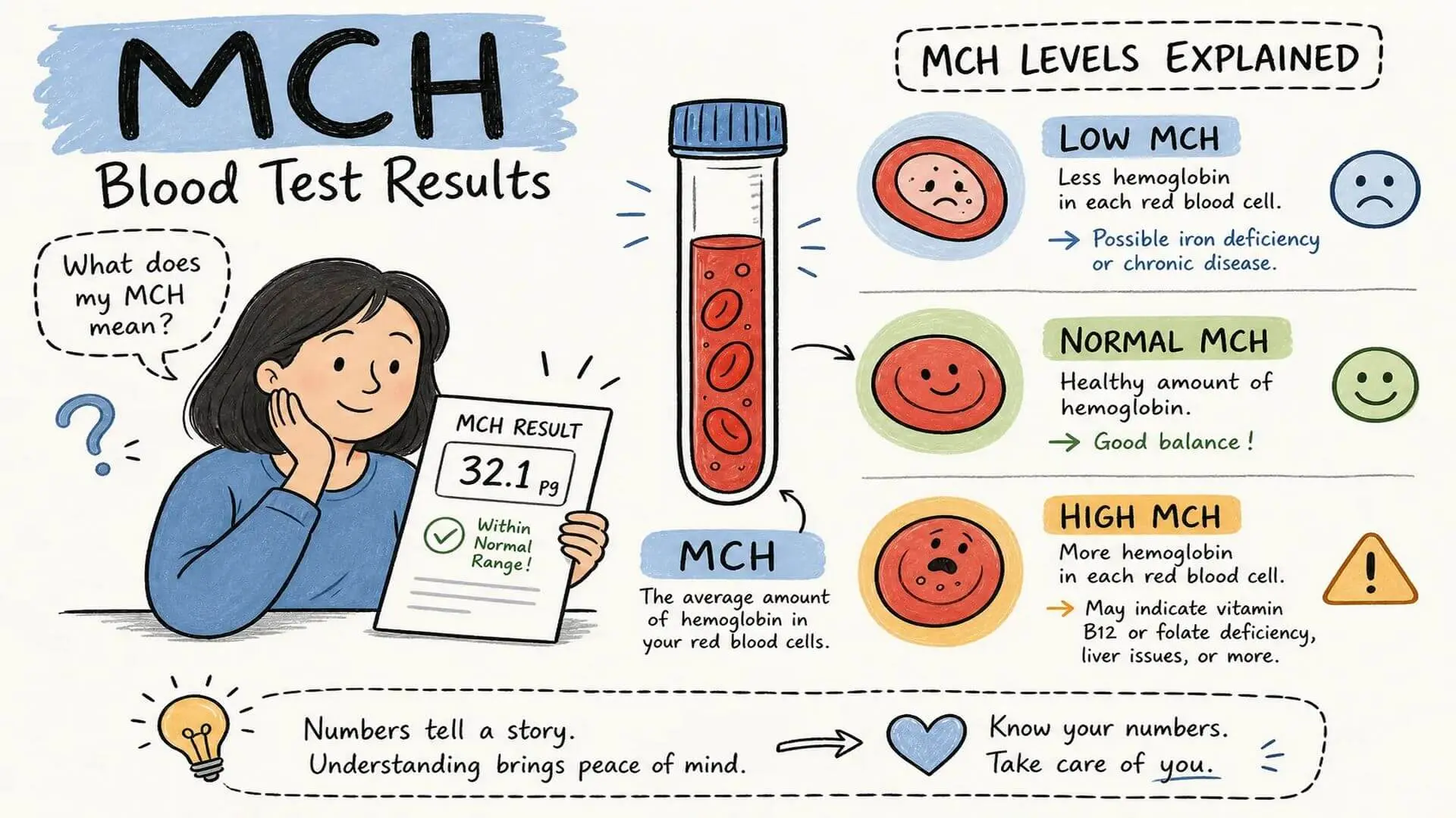
If you’ve ever looked at a CBC report and wondered what is MCH in a blood test, you’re not alone. MCH stands for mean corpuscular hemoglobin, and it measures the average amount of hemoglobin packed into a single red blood cell. Hemoglobin is the protein that carries oxygen through your body, so this value is essentially telling you how well-equipped each red blood cell is to do its job.
MCH rarely shows up as the only abnormal value on a report. It almost always appears alongside other CBC indices that, together, point your doctor toward a specific diagnosis. If you want to go deeper on related markers after reading this, related guides in this series cover MCV, MCHC, hemoglobin, and ferritin with the same evidence-first approach you’ll find here.
This article walks through what MCH measures, what normal adult values look like, what high and low results indicate, and what your next steps should be if your number falls outside the reference range.
What is MCH in a blood test, and what does it actually measure?
MCH is calculated by dividing the total hemoglobin in a blood sample by the total number of red blood cells, then expressing the result in picograms (pg). Because this calculation depends on both the amount of hemoglobin present and the size of the cells carrying it, MCH almost always moves in the same direction as MCV (mean corpuscular volume), the index that measures cell size. Larger cells tend to carry more hemoglobin, so when MCV rises, MCH typically rises with it.
This relationship is why MCH rarely tells a complete story on its own. Clinicians read it as part of a trio alongside MCV and MCHC (mean corpuscular hemoglobin concentration), using the combination to classify what type of anemia is present and narrow down its cause. That trio is covered in detail in its own section below.
MCH is commonly included on standard automated CBC panels and is frequently ordered during annual physicals, pre-surgery workups, pregnancy monitoring, and evaluations of fatigue or unexplained weakness. The value becomes clinically meaningful when it falls consistently outside the reference range printed on your specific lab report.
MCH normal range: what the numbers on your report mean
For most adults, a normal MCH falls between 27 and 33 picograms per red blood cell. Some labs narrow this to 27 to 31 pg; others report it as 26 to 33 pg, depending on the automated analyzer they use and the population their reference intervals are based on. The most reliable number to use is always the reference range on your own lab report, not a general figure from a website.
Newborns start with naturally higher MCH values, typically 31 to 37 pg, which drop significantly during infancy before climbing back toward adult levels. Pregnancy introduces a dilutional effect on blood volume, which can push MCH slightly below the standard adult threshold without indicating a disease process. Older adults generally stay within the adult range, though comorbidities such as chronic disease and B12 deficiency become more prevalent with age and can nudge values in either direction.
Sex does not significantly shift the MCH reference range. Most clinical guidelines apply the same 27 to 33 pg window to both men and women, though some labs do report slightly different intervals by sex. Your lab’s printed range is the one that matters for interpreting your specific result.
What low MCH signals about your red blood cells
A low MCH, generally below 27 pg per cell, means each red blood cell is carrying less hemoglobin than expected. In clinical practice, this almost always points to insufficient iron for hemoglobin synthesis, an inherited hemoglobin disorder, or chronic inflammation disrupting iron metabolism.
Iron deficiency: the most common cause
Iron deficiency anemia is the single most common cause of low MCH worldwide. Without adequate iron, hemoglobin synthesis stalls, producing red blood cells that are both smaller and paler than normal. Common triggers include heavy menstrual bleeding, gastrointestinal bleeding from ulcers or polyps, poor dietary intake, malabsorption conditions like celiac disease, and the increased iron demands of pregnancy.
Thalassemia is the second major cause. This inherited disorder produces defective hemoglobin chains, resulting in persistently low MCH that doesn’t respond to iron supplementation. Treating thalassemia as iron deficiency with high-dose iron can worsen iron overload rather than relieve symptoms, an important distinction that underscores why correct diagnosis matters. Anemia of chronic disease, driven by long-term inflammation from conditions like rheumatoid arthritis, kidney disease, or cancer, is another frequent driver of low MCH. Less common causes include lead poisoning and sideroblastic anemia, a bone marrow disorder that prevents the incorporation of iron into hemoglobin.
Symptoms that accompany low MCH
Fatigue and weakness are the most frequently reported symptoms. You may also notice shortness of breath during routine activity, pale skin, cold hands and feet, dizziness, or persistent headaches. In iron deficiency specifically, brittle nails and unusual cravings for non-food items (known as pica) can appear. Some people with mildly low MCH have no symptoms at all and only discover the result through routine testing, which is exactly why the CBC is a standard part of annual checkups.
What high MCH tells your doctor
A high MCH, generally above 33 pg per cell (exact cutoffs vary by lab, so check your report’s printed reference range), means each red blood cell contains more hemoglobin than normal. This almost always happens because the cells themselves are abnormally large, a condition called macrocytosis. The anemia that results from large, hemoglobin-heavy cells is classified as macrocytic anemia, and its two most common drivers are nutritional deficiencies that most people can correct with targeted treatment.
Vitamin B12 and folate deficiency as the primary drivers
Both vitamin B12 and folate (B9) are essential for normal DNA synthesis in red blood cell precursors. When either nutrient is lacking, cells divide improperly and grow oversized, carrying excess hemoglobin. B12 deficiency can arise from pernicious anemia (an autoimmune condition that blocks B12 absorption), strict vegan or vegetarian diets without supplementation, long-term metformin use, or post-surgical changes to the stomach. Folate deficiency often links to poor dietary intake, pregnancy, or medications like methotrexate that interfere with folate metabolism.
Because alcohol is directly toxic to bone marrow and disrupts folate absorption, elevated MCH can appear even in people without obvious nutritional deficiency, making chronic alcohol use one of the leading non-nutritional causes. Liver disease alters the lipid composition of red cell membranes, increasing cell size. Hypothyroidism slows bone marrow activity and can raise MCH before classic thyroid symptoms become obvious. Certain medications are also known to elevate MCH as a recognized side effect, including specific antiretrovirals, anticonvulsants such as phenytoin and valproate, and chemotherapy agents like hydroxyurea.
Neurological symptoms that distinguish B12-driven high MCH
High MCH from B12 deficiency carries neurological consequences that other causes of elevated MCH typically do not. Numbness or tingling in the hands and feet (often in a symmetrical stocking-glove pattern), difficulty with balance, memory problems, and mood changes are characteristic. Jaundice or a yellowish skin tone may also appear. These neurological symptoms can become permanent if the deficiency progresses without treatment, because B12 deficiency causes structural damage to the posterior and lateral spinal cord tracts over time. If you are experiencing any of these neurological changes alongside an elevated MCH result, that combination warrants prompt evaluation rather than a routine scheduled appointment.
Reading MCH alongside MCV and MCHC: the full picture
MCH does not exist in isolation. Clinicians read it alongside MCV, which measures red blood cell size, and MCHC, which measures the concentration of hemoglobin within a cell. Understanding how these three indices interact is what allows a CBC to point toward a specific diagnosis rather than simply flagging “anemia” as a general finding.
MCH is derived by dividing total hemoglobin by the red blood cell count. MCHC goes further by dividing hemoglobin by hematocrit, effectively controlling for cell size. Because MCH depends on cell volume, it almost always tracks in parallel with MCV. MCHC is the more sensitive marker for detecting hypochromia (pale, hemoglobin-poor cells) because it corrects for size differences that can mask the true hemoglobin concentration.
The diagnostic patterns clinicians use to classify anemia based on these three values are:
- Low MCV + low MCH + low MCHC: Microcytic hypochromic anemia, pointing to iron deficiency or thalassemia.
- High MCV + high MCH + normal MCHC: Macrocytic normochromic anemia, pointing to B12 or folate deficiency.
- Normal MCV + normal MCH + normal MCHC: Normocytic anemia, associated with acute blood loss, hemolytic anemia, or kidney failure.
- Normal or low MCV + high MCHC: Suggests hereditary spherocytosis, where cells are spherical and densely packed with hemoglobin.
For readers who want to explore MCV and MCHC in greater depth, related guides in this series cover each index with full clinical context, including how RDW helps distinguish iron deficiency from thalassemia when MCV and MCH look similar.
What to do after an abnormal MCH result
An abnormal MCH on its own is a prompt to investigate further, not a diagnosis. The follow-up process is structured and most underlying causes are treatable once identified, whether your number is low and pointing toward iron stores, or high and suggesting a vitamin deficiency or other driver.
Tests your doctor is likely to order
When MCH is low, the standard first step is a complete iron panel: serum ferritin, serum iron, and transferrin saturation. Low ferritin (below 30 ng/mL) combined with low transferrin saturation (below 16%) is consistent with iron deficiency, though it’s worth noting that ferritin is also an acute-phase reactant and can appear falsely normal or elevated in the setting of inflammation, a nuance your doctor will factor in. If ferritin is normal but MCH and MCV remain persistently low, hemoglobin electrophoresis is ordered to evaluate for thalassemia. A peripheral blood smear may be added to visualize cell shape and color directly.
For high MCH, serum B12 and folate levels are the standard first tests. These are typically followed by a reticulocyte count and, if needed, a peripheral blood smear. Thyroid function tests (TSH) and liver function tests are added when hypothyroidism or liver disease is on the differential. If a medication side effect is suspected, the prescribing physician will review whether the drug is a known cause of macrocytosis.
Symptoms that require urgent attention
Most abnormal MCH results can be addressed through a scheduled appointment with your primary care provider. However, some accompanying symptoms indicate that waiting is not the right approach. Seek prompt evaluation if you experience chest pain, severe shortness of breath at rest, rapid or irregular heartbeat, fainting, black or bloody stools, or unexplained weight loss alongside an abnormal MCH. Neurological changes, including worsening numbness, inability to walk steadily, or sudden confusion, are also red-flag symptoms, particularly when high MCH points toward B12 deficiency as the cause.
Key takeaways: what your MCH result is actually telling you
So what is MCH in a blood test, and what should you do with the number? At its core, MCH tells you how much hemoglobin, on average, each of your red blood cells is carrying. Normal adult values generally fall between 27 and 33 pg, though your lab’s printed range is the one to apply to your specific result. When MCH falls low, the investigation typically starts with iron stores; when it runs high, B12 and folate are usually checked first.
Here’s what to do next: bring your full CBC report to your appointment, ask your doctor which indices are outside the reference range and why, and request a targeted follow-up test rather than a generalized recheck in three months. The sooner the underlying cause is identified, the sooner it can be addressed. For a deeper look at related markers, including ferritin, hemoglobin, MCV, and HbA1c, explore BioScience’s blood biomarker guide series, where each index receives the same evidence-first, expert-reviewed treatment you found here.
The information on this page is peer reviewed by a qualified editorial review board member. Learn more about us and our editorial process.
Last reviewed on .
Article history
- Latest version
Reference(s)
- Cleveland Clinic. “Mean Corpuscular Hemoglobin (MCH).” Cleveland Clinic <https://my.clevelandclinic.org/health/diagnostics/mch-blood-test>.
- Voukelatou, Panagiota., et al. “Neurologic symptoms as the only manifestation of B12 deficiency in a young patient with normal hematocrit, MCV, peripheral blood smear and homocysteine levels.” Oxford Medical Case Reports, vol. 2016, no. 12, 2016, doi: 10.1093/omcr/omw091. <https://pmc.ncbi.nlm.nih.gov/articles/PMC5184828/>.
- Walker, H. Kenneth. “Clinical Methods: The History, Physical, and Laboratory Examinations.” 3rd ed., Butterworth-Heinemann, 1990, isbn: 9780409900774. <https://www.ncbi.nlm.nih.gov/books/NBK260/>.
Cite this page:
- Posted by Dayyal Dungrela