Wright’s Stain: Preparation, Principle, Procedure, and Results
Learn how to prepare, apply, and interpret Wright’s stain for accurate diagnosis and monitoring of blood-related disorders.
Wright’s stain is a Romanowsky-type stain developed by James Homer Wright in 1902. It combines eosin and methylene blue dyes to differentiate blood cells, highlighting cellular structures in blood and bone marrow smears. Commonly used in hematology, it aids in diagnosing infections, blood disorders, and morphological abnormalities. Unlike Wright-Giemsa stain, it is simpler yet highly effective for routine diagnostics.
Principle of Wright’s Stain
The principle of Wright’s stain relies on differential staining using a combination of acidic and basic dyes, enabling precise visualization of blood cell types and structures. Eosin, an acidic anionic dye, adheres to basic cellular components like hemoglobin and eosinophilic granules, giving them shades of red, pink, or orange. Conversely, methylene blue, a basic cationic dye, interacts with acidic structures such as nuclei and basophilic granules, coloring them blue or purple.
When diluted with buffered water, this polychromatic staining method triggers ionization, allowing the dyes to bind specifically to cellular components. Neutral elements are stained by both dyes, producing a range of variable colors. The methanol-based composition of Wright’s stain eliminates the need for a separate fixation step. However, fixation with methanol is recommended to reduce water artifacts, particularly on humid days or when using aged stain.
Wright Stain Preparation
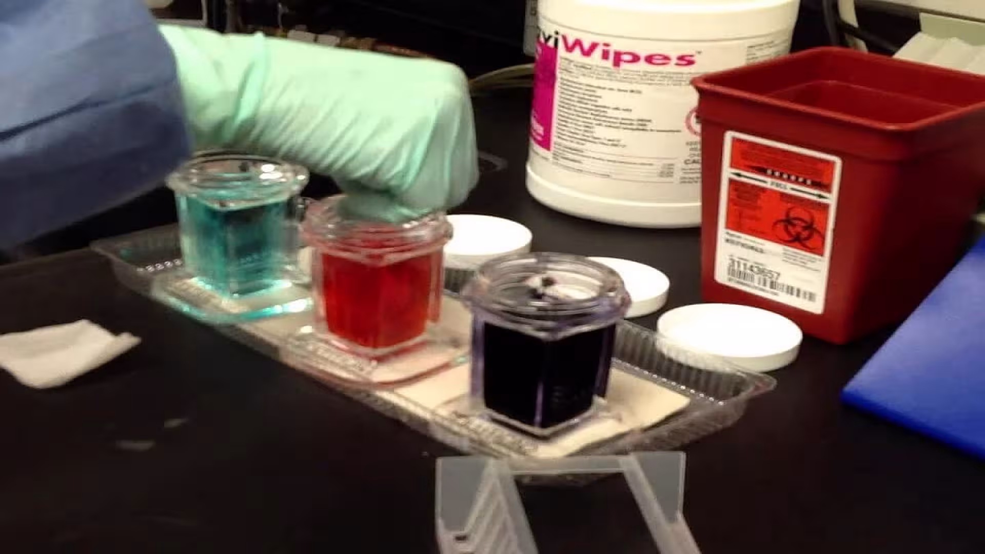
Dissolve 0.1 g dry Wright’s stain (eosin methylene blue) in 60 ml methanol. Grind eosin Y into a fine powder using a mortar and pestle. Add 10 ml methanol to dissolve the powder, then gradually mix in the remaining methanol. Transfer the solution to a clean storage bottle, repeating the process with methanol until all the stain is fully dissolved. Store the solution in a sealed, dark container at room temperature.
Wright Stain Procedure
Prepare a blood smear by spreading a drop of blood on a clean glass slide and air-drying it. Flood the slide with Wright stain solution and let it stand for 1–3 minutes. Add an equal volume of phosphate buffer (pH 6.4–6.8) and gently mix by tilting the slide. Incubate for 5–7 minutes, rinse with distilled water to remove excess stain, and air-dry before microscopic examination.
Results and Interpretation of Wright’s Stain
| Cell Type | Staining Characteristics |
|---|---|
| Erythrocytes | Yellowish-red |
| Neutrophils | Nucleus: Dark purple Granules: Reddish-lilac Cytoplasm: Pale pink |
| Eosinophils | Nucleus: Blue Granules: Red to orange-red Cytoplasm: Blue |
| Basophils | Nucleus: Purple to dark blue Granules: Dark purple |
| Lymphocytes | Nucleus: Dark purple Cytoplasm: Sky blue |
| Monocytes | Nucleus: Dark purple Cytoplasm: Mosaic pink and blue |
| Platelets | Violet to purple granules |
Normal Wright Stain Results
- Red Blood Cells (RBCs): Appear pink to red with a uniform circular shape.
- White Blood Cells (WBCs): Nuclei stain purple, while cytoplasm varies based on cell type.
- Platelets: Appear as small purple granules scattered throughout the smear.
Abnormal Findings in Wright Stain
- Anemia: Altered RBC morphology, such as anisocytosis or poikilocytosis.
- Leukemia: Abnormal WBC shapes and increased counts.
- Infections: Presence of toxic granulations in neutrophils.
Advantages and Limitations of Wright’s Stain
Advantages
- Provides clear differentiation of cellular components.
- Rapid and reliable results.
- Essential for diagnosing blood disorders.
Limitations
- Requires precise preparation and technique.
- Artifacts may form if improperly prepared.
- Limited use in identifying subtle chromosomal abnormalities.
Wright Stain vs. Giemsa Stain
| Feature | Wright Stain | Giemsa Stain |
|---|---|---|
| Composition | Eosin and methylene blue | Eosin, methylene blue, and azure dyes |
| Application | Routine blood smear analysis | Malaria and parasite identification |
| Staining Time | Shorter | Longer |
Wright-Giemsa Stain Procedure Differences
The Wright-Giemsa stain involves additional steps, such as prolonged staining time, to enhance visualization of intracellular structures.
Conclusion
Wright’s stain is an essential technique in hematology, providing clear differentiation of blood cells for accurate diagnosis and monitoring of blood-related disorders. Its ability to highlight cellular structures with precision makes it a fundamental tool in clinical and research laboratories. By adhering to proper preparation and procedures, laboratory professionals can ensure reliable and consistent results.
Widely used for routine blood smears and specialized applications, Wright’s stain remains a gold standard in hematological diagnostics. Its simplicity and reliability contribute to accurate analysis, ultimately supporting optimal patient care.
FAQs
What is the principle of Wright stain procedure?
How to perform modified Wright stain?
What is the difference between Wright stain and Wright-Giemsa stain?
What are common issues in Wright stain preparation?
The information on this page is peer reviewed by a qualified editorial review board member. Learn more about us and our editorial process.
Last reviewed on .
Article history
- Latest version
Cite this page:
- Posted by Dayyal Dungrela