White Blood Cells Morphology
Explore the detailed morphology of normal and abnormal white blood cells, including key characteristics of WBC morphology abnormalities crucial for diagnosis.

To accurately assess the total leukocyte count, examining a blood smear under a high-power objective (40× or 45×) is a valuable initial step. Following this, it is essential to perform a differential leukocyte count to identify and categorize the various types of white blood cells. Abnormally appearing leukocytes require further evaluation under an oil-immersion objective to ensure a precise diagnosis.
Morphology of Normal Leukocytes
Box 1: Role of blood smear in leukemia
- It suggests the diagnosis or differential diagnosis.
- It suggests further investigations to be performed for definitive diagnosis.
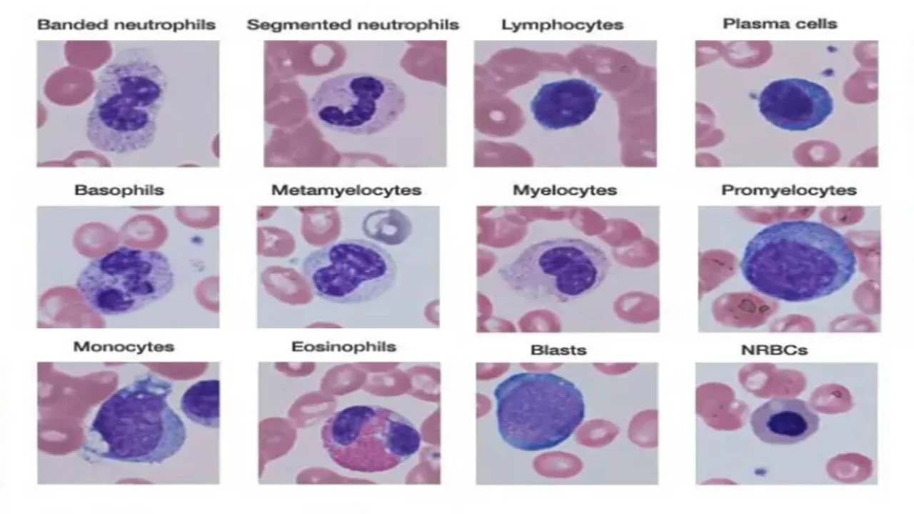
The morphology of normal leukocytes can be observed in a standard blood smear, as illustrated in Figure 1. A blood smear plays a critical role in diagnosing and differentiating various hematological disorders, such as leukemia. Specifically, it can guide the clinician in deciding on further investigations necessary for a definitive diagnosis.
- Polymorphonuclear Neutrophils: These neutrophils are typically 14-15 μm in diameter. The cytoplasm appears colorless or slightly eosinophilic, containing numerous small, fine mauve granules. The nucleus of a neutrophil is segmented, with 2-5 lobes connected by delicate chromatin strands, and the chromatin itself is dense and stains deep purple. Band neutrophils, which are a precursor to segmented neutrophils, have a U-shaped, non-segmented nucleus. Normally, band neutrophils make up less than 3% of all leukocytes. Among segmented neutrophils, the majority possess three lobes, with fewer than 5% exhibiting five lobes. Additionally, in females, 2-3% of neutrophils display a small nuclear projection, known as a drumstick, representing an inactivated X chromosome.
- Eosinophils: Slightly larger than neutrophils, eosinophils measure around 15-16 μm. Their cytoplasm is densely packed with large, bright orange-red granules, and their nucleus is typically bilobed. Eosinophils are often seen partially ruptured in blood smears.
- Basophils: Basophils, which are infrequently observed on normal blood smears, are smaller cells (9-12 μm) with large, coarse, deep purple granules. These granules often obscure the nucleus, making it challenging to discern.
- Monocytes: The largest of the leukocytes, monocytes range from 15-20 μm in size. These cells are irregularly shaped with an oval or kidney-shaped nucleus and fine, delicate chromatin. Their abundant cytoplasm is blue-gray with a ground glass appearance, often containing fine azurophilic granules and vacuoles. Once monocytes migrate from the bloodstream into tissues, they differentiate into macrophages.
- Lymphocytes: Two types of lymphocytes are identifiable in peripheral blood smears: small and large. Small lymphocytes, which predominate, measure 7-8 μm and have a high nuclear-cytoplasmic ratio, with a thin rim of deep blue cytoplasm. The nucleus is round or slightly clefted, with coarsely clumped chromatin. In contrast, large lymphocytes (10-15 μm) have a more abundant, pale blue cytoplasm that may contain a few azurophilic granules, and their nucleus is often oval or round, typically located on one side of the cell.

Morphology of Abnormal Leukocytes
Abnormalities in leukocyte morphology often indicate underlying pathological conditions and are critical for diagnosis.
- Toxic Granules: These are intensely stained blue-purple, coarse granules within the cytoplasm of neutrophils, commonly observed in severe bacterial infections.
- Döhle Inclusion Bodies: Döhle bodies are small, oval, pale blue cytoplasmic inclusions found in the periphery of neutrophils, representing remnants of ribosomes and rough endoplasmic reticulum. They are often present alongside toxic granules in bacterial infections.
- Cytoplasmic Vacuoles: The presence of vacuoles in neutrophils typically indicates phagocytosis and is frequently seen in bacterial infections.
- Shift to the Left of Neutrophils: This term refers to the appearance of immature neutrophil forms (such as band forms and metamyelocytes) in the peripheral blood, commonly associated with infections and inflammatory disorders.
- Hypersegmented Neutrophils: Hypersegmentation occurs when more than 5% of neutrophils exhibit five or more nuclear lobes. These cells, also known as macropolycytes, are typically larger and are an early indicator of folate or vitamin B12 deficiency.
- Pelger-Huet Cells: In the Pelger-Huet anomaly, a benign autosomal dominant condition, granulocytes fail to undergo normal nuclear segmentation, resulting in rod-like, round, or bilobed nuclei. Similar cells, termed pseudo-Pelger-Huet cells, are also observed in myeloproliferative disorders.
- Atypical Lymphocytes: Atypical lymphocytes, which are large and irregularly shaped with abundant cytoplasm and irregular nuclei, are frequently seen in viral infections, particularly infectious mononucleosis. The cytoplasm of these cells shows deep basophilia at the edges, with scalloped borders, and the nuclear chromatin is less condensed, occasionally containing a nucleolus.
- Blast Cells: These are the most immature forms of leukocytes, measuring 15-25 μm in diameter. They are characterized by a high nuclear-cytoplasmic ratio, with one or more nucleoli and immature nuclear chromatin. Blast cells are typically seen in severe infections, infiltrative disorders, and leukemia. In cases of leukemia and lymphoma, the blood smear plays a crucial role in suggesting the diagnosis and guiding further diagnostic tests.

The information on this page is peer reviewed by a qualified editorial review board member. Learn more about us and our editorial process.
Last reviewed on .
Article history
- Latest version
Cite this page:
- Posted by Dayyal Dungrela