Comparative Anatomy: Fish Brain Vs. Frog Brain
Discover the key differences between fish and frog brains. Learn how the structure of the nervous system varies in fish and frogs, including detailed brain anatomy.
The nervous system, originating from ectodermal tissue, forms a highly sensitive and interconnected system in vertebrates. The brain, positioned at the anterior end of the central nervous system, is a universal structure among bilaterally symmetrical vertebrates, sharing a common architectural foundation. Although the general plan of the brain is consistent across species, the brain of lower vertebrates, such as fish and frogs, varies in structure and function based on evolutionary adaptations.
In its early embryonic stages, and in lower vertebrates, the brain appears as the swollen anterior portion of the neural tube, commonly referred to as the "encephalon." This structure undergoes differential growth, resulting in the division into three primary cerebral vesicles: the forebrain (prosencephalon), midbrain (mesencephalon), and hindbrain (rhombencephalon). These divisions are key to understanding brain development and specialization across vertebrates.
Brain Differentiation in Vertebrates
The brain of various vertebrate groups, particularly fish and frogs, demonstrates important evolutionary modifications. In lower vertebrates, such as fish, the olfactory lobes are highly developed, emphasizing the reliance on smell for survival. However, as vertebrates ascend the evolutionary ladder, these olfactory lobes reduce in prominence, particularly in amphibians like frogs. The overall structure of the brain becomes more complex, with increased segregation of the brain into specific regions responsible for distinct functions.
In terms of ventricle structure, the brain is divided into four ventricles: the lateral ventricles (1 and 2, also known as paracoels), the third ventricle (diacoel), and the fourth ventricle (myelocoel). Communication between the lateral ventricles and the third ventricle occurs through an aperture called the foramen of Monro, while the third and fourth ventricles are connected via the cerebral aqueduct (also known as the Aqueduct of Sylvius).

Structural Composition of the Brain
The brain's tissue is categorized into grey matter and white matter. Grey matter primarily consists of nerve cell bodies, dendrites, and proximal axons, while white matter comprises medullated fibers responsible for conducting impulses between different parts of the brain and the spinal cord. Within the ventricles and surrounding spaces, cerebrospinal fluid circulates, playing a protective and nourishing role. This fluid, produced by the choroid plexus, is also present between the brain's meninges and is reabsorbed into the blood vessels.


Comparative Anatomy: Fish Brain vs. Frog Brain
When comparing the brains of fish and frogs, several anatomical differences become apparent. For example, in fish (Scoliodon), the brain is enclosed within a cartilaginous cranium, whereas the frog brain resides within a bony cranium. Moreover, fish possess a single membrane known as meninx primitiva that covers the brain, whereas frogs have two protective meninges: an outer dura mater and an inner pia mater.
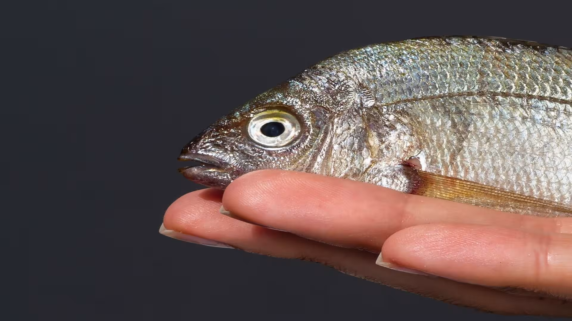
Fish exhibit highly developed olfactory lobes, reflecting their dependence on the sense of smell for hunting and survival. In contrast, frogs possess smaller olfactory lobes, indicating a lesser reliance on smell. Furthermore, the brain ventricles in fish are spacious, while in frogs, they are comparatively narrow. The cerebrum in fish is typically rectangular and undivided, whereas frogs display more advanced brain structures, with their cerebrum divided into two hemispheres.
Another key difference lies in the development of the cerebral cortex. In fish, the grey matter is confined to the lining of the lateral ventricles, with no defined cerebral cortex. Conversely, frogs exhibit a more developed cortex, with some nerve cells migrating to the brain’s surface to form this crucial structure. Additionally, the diencephalon in fish is short and narrow, while it is wider and more rhomboidal in frogs, highlighting evolutionary adaptations to different ecological roles.
The midbrain (mesencephalon) and hindbrain (rhombencephalon) also exhibit structural differences between fish and frogs. For instance, the optic lobes in fish are largely covered by the cerebellum, while in frogs, these lobes are more exposed. In fish, the cerebellum is large and divided into lobes, facilitating complex motor functions, whereas in frogs, it is smaller and undivided, reflecting less need for intricate motor coordination.
| Fish (Shark: Scoliodon laticaudus) Brain | Frog Brain |
|---|---|
| The brain lies in a cartilaginous cranium. | The brain lies in a bony cranium. |
| A single outer membrane, 'Meninge primitiva,' covers the brain. | An outer thick dura mater and inner thinner pia mater protect the brain, resulting in two meninges (membranes). |
| All the ventricles in the brain are spacious. | All the ventricles in the brain are relatively small and narrow. |
| Prosencephalon | |
| The olfactory lobes are highly developed, aiding in a strong reliance on smell to detect prey. | The olfactory lobes are relatively small, aiding in a less significant reliance on smell. |
| The olfactory lobes extend forward and outward from the anterolateral angles of the cerebrum, lying side by side. | The olfactory lobes lie side by side without the differentiation seen in sharks. |
| Each olfactory lobe differentiates into an olfactory tract and a bilobed olfactory bulb. The olfactory bulbs are closely applied to the olfactory sacs. | No such close contact exists in the frog brain. |
| The cerebrum is somewhat rectangular, large, and undivided. | The cerebrum is divided into two ovoid cerebral hemispheres by a median fissure. |
| A median neuropore is present on the ventral side of the cerebrum for terminal nerves. | There is no neuropore in the frog brain. |
| Pallium and corpora striata are poorly developed. | Pallium and corpora striata are well developed. |
| There is no cerebral cortex; the grey matter is restricted to the lining of the lateral ventricles. | Some of the nerve cells from the inner grey matter migrate to the surface to form the cerebral cortex. |
| The diencephalon is short and narrow, almost fully covered dorsally by the cerebrum. | The diencephalon is wide and rhomboidal, covered either dorsally or ventrally by the cerebrum. |
| The pineal stalk is long and slender, extending forward and upward. | The pineal stalk is very short, bearing a knob-like pineal body distally (in tadpoles). |
| The infundibulum consists of a large median lobe and a pair of small lateral lobes called 'Lobi inferiores'. | The infundibulum is a median bilobed structure. |
| The median lobe of the infundibulum and the hypophysis form the 'pituitary body'. | The entire infundibulum and hypophysis form the pituitary body. |
| A pair of hollow outgrowths, 'Sacci vasculosi,' are present on the sides of the hypophysis. These act as pressure receptors. | Sacci vasculosi are absent in the frog brain. |
| Mesencephalon | |
| The optic lobes are largely covered by the cerebellum. | The optic lobes are not covered by any brain structure. |
| The crura cerebri are largely covered by the lobi interiores and sacci vasculosi. | The crura cerebri are slightly covered by the pituitary body. |
| Rhombencephalon | |
| The cerebellum is a large rhomboidal structure divided into three lobes by two deep transverse furrows. | The cerebellum is a small transverse band, undivided, and does not cover any part of the brain. |
| A pair of irregular, thin-walled sacs called 'Corpora restiformia' are present on the sides of the anterior cerebellum. | Corpora restiformia are absent in the frog brain. |
| The medulla oblongata is partly covered in front by the cerebellum. | The medulla oblongata is not covered by the cerebellum. |
| There are three commissures: the 'anterior' in lamina terminalis, the 'dorsal' in front of the pineal stalk, and the 'posterior' between the diencephalon and optic lobes. | There are four commissures: the 'anterior' in lamina terminalis, the 'hippocampal' above the anterior commissure, the 'dorsal' in front of the pineal stalk, and the 'posterior' between the diencephalon and optic lobes. |
| The dorsal commissure is more developed, while the posterior commissure is less developed than in frogs. | The dorsal commissure is less developed, while the posterior commissure is more prominent than in sharks. |
Functional Roles of Brain Regions in Frogs
- Olfactory lobes: Responsible for the sense of smell, though less developed than in fish.
- Cerebral hemispheres: The seat of intelligence and memory, facilitating more complex behaviors.
- Diencephalon: Governs metabolic processes and maintains homeostasis.
- Optic lobes: Essential for vision and processing visual stimuli.
- Cerebellum: Coordinates voluntary muscle movements.
- Medulla oblongata: Regulates involuntary functions, such as breathing and heart rate.
FAQs
Do fish have brains?
What is the structure of a fish brain?
Do fish have a brain similar to mammals?
Does a frog have a brain?
How does a frog brain differ from a fish brain?
What is the frog dorsal brain model?
What functions do the olfactory lobes serve in fish and frogs?
How do the nervous systems of fish and frogs compare?
Reference(s)
- R.L. Kotpal. Modern Text Book Of Zoology: Vertebrates. Rastogi Publications. 5thEdition. 1 January 2022. ISBN: 9788193887561.
- Stephen Miller and John Harley. Zoology. McGraw-Hill Professional. 8thEdition. 16 October 2009. ISBN: 9780070164833.
Cite this page:
- Posted by Dr. Nida Hayat Khan