Gram Staining: Purpose, Principle, Procedure and Observation
Learn about the Gram stain technique for classifying Gram-positive and Gram-negative bacteria. Discover the process, reagents, and significance of Gram staining in bacterial identification and differentiation.
In 1883, Dr. Hans Christian Gram (1853-1938) pioneered a technique for categorizing bacteria into two major groups: Gram-positive and Gram-negative. Initially published in 1884, this method remains the most pivotal staining technique for bacterial classification and differentiation.
The Gram stain procedure involves four key reagents: crystal violet (primary dye), Gram's iodine (mordant), ethyl alcohol (decolorizer), and safranin (counterstain). Gram-positive bacteria retain the primary dye (crystal violet) even after the decolorization process, while Gram-negative bacteria lose the primary dye and subsequently take up the color of the counterstain (safranin).
The Gram reaction depends on the chemical composition of the bacterial cell wall, particularly its lipid content. Gram-negative cell walls contain 11-22% lipids, whereas Gram-positive cell walls contain only 1-4%. In Gram-negative cells, the high lipid content, when exposed to alcohol, leads to the formation of large pores in the cell wall. The dehydrating effect of alcohol cannot seal these pores, resulting in the loss of the primary stain and leaving the cell colorless. These cells then take up the counterstain (safranin). Conversely, the low lipid content in Gram-positive cell walls forms small pores when dissolved in alcohol. The dehydrating effect of alcohol closes these pores, preventing the primary dye (crystal violet) from leaving the cell.
Purpose
The purpose of the Gram stain technique is the identification, differentiation, and classification of bacteria.
Needs
Specimen
- Sputum
- Body fluid
- Pus
- Swab of cells from the infection site
- Sample of bacteria or fungi grown and isolated in culture
Reagents
- Crystal violet
- Gram iodine
- Ethyl alcohol (95%)
- Safranin
- Cedar wood oil
Equipment
- Bunsen burner
- Wire loop
- Glass slides
- Microscope
Procedure
- Prepare a smear of the specimen, air dry it, and then fix it with a low flame.
- Flood the smear with crystal violet and let it sit for 1 minute. Rinse the smear with running tap water.
- Pour Gram's iodine on the smear, let it sit for 1 minute, then rinse with tap water.
- Decolorize the smear by pouring alcohol until the purple color no longer washes out.
- Apply safranin to the smear and let it sit for 45 seconds, then rinse with tap water and air dry.
- Examine the stained smear under an oil immersion lens and note the arrangement, shape, and Gram reaction of the cells.
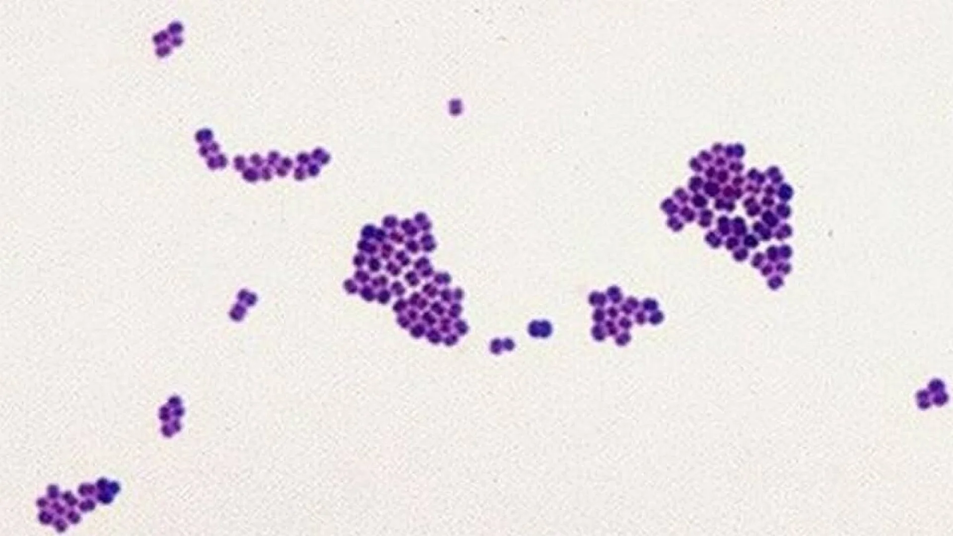
Observation
Gram-positive bacteria will appear purple, while Gram-negative bacteria will appear pink.
The information on this page is peer reviewed by a qualified editorial review board member. Learn more about us and our editorial process.
Last reviewed on .
Article history
- Latest version
Cite this page:
- Comment
- Posted by Dayyal Dungrela