May Grünwald-Giemsa Stain: Principle, Procedure, and Protocol
Learn about the MGG stain principle, procedure, and protocol. Explore the step-by-step MGG stain procedure for accurate blood and bone marrow analysis.
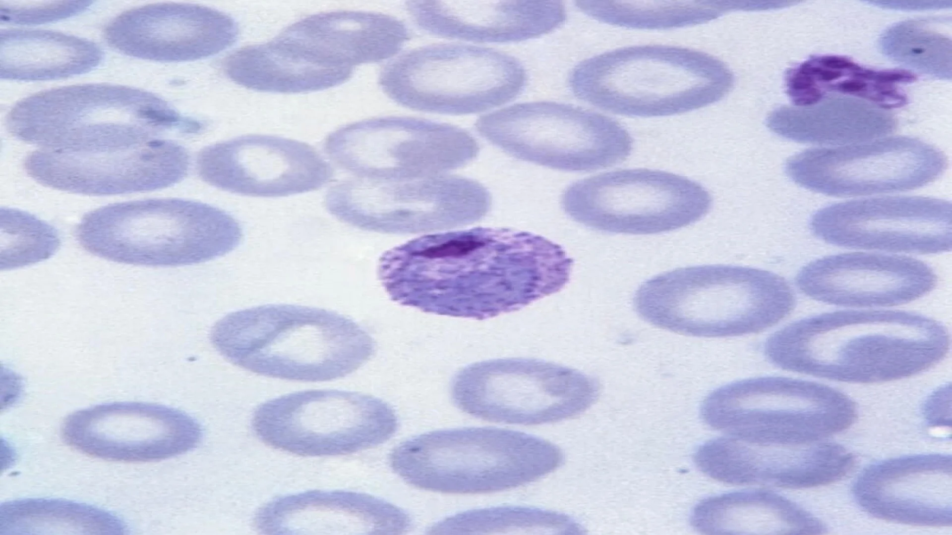
The May-Grünwald-Giemsa (MGG) stain is a Romanowsky-type stain widely used in hematology to differentiate and highlight blood cells, including neutrophils, eosinophils, basophils, lymphocytes, and monocytes. It is essential for examining blood smears, bone marrow aspirates, and tissue samples, providing clear cellular morphology for diagnostic purposes.
Principle of MGG Stain
May-Grünwald Giemsa (MGG) staining uses acidic eosin and basic dyes like methylene blue and azure B to differentiate cell structures. Eosin stains cytoplasm pink/red, while basic dyes stain nuclei blue/purple. This dual staining relies on precise pH for accurate visualization of blood and bone marrow cells.
Components of the May-Grünwald Giemsa (MGG) Stain
The May-Grünwald Giemsa (MGG) stain consists of:
- May-Grünwald Solution: Methylene blue and eosin dissolved in methanol.
- Giemsa Solution: Methylene blue, azure B, and eosin in glycerol and methanol.
- Buffer Solution: Ensures optimal pH (typically 6.8) for accurate staining results.
Procedure and Protocol
The May-Grünwald Giemsa (MGG) stain procedure is as follows:
- Prepare Sample: Make a thin smear of blood or bone marrow on a glass slide and air-dry.
- Fixation: Immerse the slide in methanol for 3-5 minutes to fix cells.
- May-Grünwald Staining: Apply May-Grünwald solution for 3 minutes, then add an equal volume of buffer solution and leave for 3 minutes. Rinse with buffer.
- Giemsa Staining: Stain with Giemsa solution (1:9 dilution with buffer) for 10-15 minutes.
- Rinsing: Wash with buffered water to remove excess stain.
- Drying: Air-dry the slide upright.
- Examination: Observe under a microscope, starting with low magnification.
May-Grünwald Giemsa Stain for Bone Marrow Examination
May-Grünwald Giemsa (MGG) stain is a two-step procedure used to analyze bone marrow cells.
- Prepare Smear: Create a concentrated, mono-layered smear of bone marrow cells on a glass slide and air-dry.
- Staining Process:
- Apply May-Grünwald solution for 3 minutes, then mix with buffer solution for another 3 minutes.
- Stain with Giemsa solution (1:9 dilution in buffer) for 10-15 minutes.
- Examination: Rinse, air-dry, and examine the slide under a microscope to evaluate cell morphology and detect hematologic abnormalities like leukemia or anemia.
May-Grünwald Giemsa Stain vs. Wright Stain
May-Grünwald Giemsa (MGG) stain and Wright stain are both Romanowsky stains but differ in intensity, time, and applications:
- Staining Intensity: MGG produces more intense staining, while Wright stain has milder color differentiation.
- Duration: MGG staining takes longer due to its two-step process, whereas Wright stain can be completed in about 30 seconds.
- Applications:
- MGG stain: Ideal for detailed morphological studies, such as bone marrow and complex blood diagnostics.
- Wright stain: Commonly used for routine blood smears and rapid assessments.
| Feature | May-Grünwald Giemsa Stain | Wright Stain |
|---|---|---|
| Detail Level | High | Moderate |
| Staining Time | Longer | Shorter |
| Cytoplasmic Detail | Detailed | Moderate |
| Application | Bone marrow, cytology | Routine blood smears |
Advantages and Limitations of the May-Grünwald Giemsa Stain
The May-Grünwald Giemsa (MGG) stain is widely used in hematology and cytology for detailed cell analysis.
Advantages:
- Superior Cellular Differentiation: Offers detailed visualization of cell morphology, including cytoplasmic features and nuclear details.
- Diagnostic Versatility: Suitable for blood and bone marrow samples, aiding in the diagnosis of hematologic conditions like leukemia and anemia.
- Reliability: Produces consistent and reproducible results in various applications.
Limitations:
- Time-Consuming: Requires a longer staining procedure compared to simpler methods.
- Preparation Sensitivity: Needs careful preparation to avoid artifacts and ensure optimal results.
- Precipitation Issues: The stain has a tendency to precipitate, which can affect staining quality if not handled properly.
- Critical pH Requirement: Staining accuracy depends on maintaining an optimal pH range of 6.5–6.8.
| Aspect | Advantage | Limitation |
|---|---|---|
| Cellular Detail | Provides superior cellular differentiation and morphological analysis. | Requires meticulous preparation to avoid artifacts. |
| Application | Suitable for blood and bone marrow analysis, aiding in the diagnosis of various hematologic conditions. | Time-consuming compared to simpler staining methods. |
| Stain Reliability | Produces consistent and reproducible results in cytological studies. | Precipitation of the stain can occur if not handled properly. |
| pH Sensitivity | Works effectively within a specific pH range (6.5–6.8) for optimal staining. | Staining can fail outside the recommended pH range, affecting results. |
| Diagnostic Versatility | Enables the detection of hematologic abnormalities like leukemia and anemia. | Longer staining time can be impractical in urgent situations. |
Summary
The May-Grünwald Giemsa (MGG) stain is a two-step procedure widely used in hematology and cytology for the differential staining of blood and bone marrow cells. This Romanowsky-type stain highlights various cell components with different affinities for acidic and basic dyes.
Features:
- Differentiates Blood Cells: The stain helps distinguish different types of white blood cells (neutrophils, eosinophils, basophils, lymphocytes, monocytes) and erythrocytes (red blood cells).
- Applications: Primarily used in hematology for blood smears and bone marrow samples, it also finds applications in histology for tissue sections.
- Precision: Offers detailed visualization of cellular morphology, aiding in the diagnosis of conditions like leukemia, anemia, and myelodysplastic syndromes.
This stain uses a combination of May-Grünwald and Giemsa solutions, which together produce a vivid contrast between cell components, making it invaluable for diagnostic and research purposes.
FAQs
What is the principle of the May-Grünwald Giemsa stain?
How is the May-Grünwald Giemsa stain used in bone marrow analysis?
What is the difference between the May-Grünwald Giemsa stain and the Wright stain?
The information on this page is peer reviewed by a qualified editorial review board member. Learn more about us and our editorial process.
Last reviewed on .
Article history
- Latest version
Cite this page:
- Posted by Dayyal Dungrela