Papanicolaou Stain (Pap Smear): Principles, Procedures, and Protocols
Learn about the Papanicolaou stain (Pap smear), a vital diagnostic tool for cervical cancer screening. Discover its principles, procedures, protocols, and applications in women’s health.
The Papanicolaou stain, or Pap stain, is a multicolored cytological staining technique developed by Dr. George Papanicolaou in 1942. It transformed cytology by allowing the early detection of cervical cancer and other abnormalities through Pap smear tests. This technique differentiates cellular components clearly and is widely used in diagnostic pathology and preventive healthcare.
What is the Papanicolaou Stain?
The Papanicolaou stain, or Pap stain, is a cytological staining technique used to differentiate cellular components under a microscope. It employs dyes such as orange G, eosin Y, and light green SF to stain cells in hues of blue (basophilia), pink (acidophilia), orange (orangeophilia), or gray-blue. Widely used in Pap smear tests, it aids in detecting cervical cancer and other cytological abnormalities.
Why is the Papanicolaou Stain Used for Cervical Cancer Screening?
The Papanicolaou stain is used for cervical cancer screening because it highlights nuclear and cytoplasmic details, allowing pathologists to detect abnormal cellular changes. This method helps identify precancerous conditions and early-stage malignancies, enabling timely treatment and improving patient outcomes.
Principles of the Papanicolaou Stain
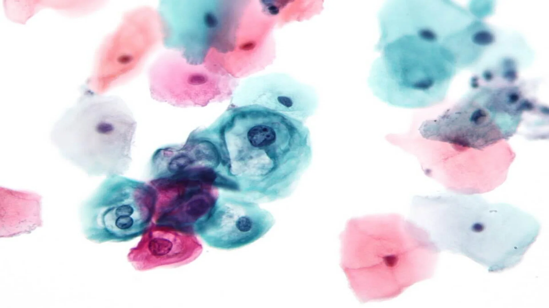
The principle of the Papanicolaou stain is to differentially stain cellular components based on their chemical and structural properties, enhancing visualization under the microscope. Hematoxylin, a basic dye, binds to acidic nuclear material, staining it blue or purple, allowing precise identification of nuclear abnormalities. Cytoplasmic components are stained with Orange G (an acid dye) and Eosin Azure (EA), a polychromatic mixture containing eosin Y, light green SF, and Bismarck brown. These dyes impart distinct shades: orange for keratin, pink for superficial squamous cells, and green for metabolically active cells like parabasal or endocervical cells.
The stain’s mechanism relies on ionic interactions, where acidic and basic dyes bind selectively to cellular components. Internal controls, such as the greenish stain in neutrophils, ensure staining quality. This method enables clear differentiation of normal, precancerous, and abnormal cells, making it essential for cytological studies like Pap smears.
Components of the Papanicolaou Stain
The Papanicolaou staining method uses a combination of dyes and reagents, each with a specific role:
- Hematoxylin: Stains nuclear material blue to purple.
- Orange G (OG): Highlights keratinized cells with an orange hue.
- Eosin Azure (EA): Differentiates cytoplasmic components, imparting pink, green, or blue shades.
Role of Each Component in Staining
- Hematoxylin: Provides contrast by emphasizing nuclear details.
- Orange G: Identifies keratinized cells, often seen in certain cancers.
- Eosin Azure: Differentiates non-keratinized cells and enhances cytoplasmic clarity.
Papanicolaou Stain Procedure
Pre-staining Preparation
- Sample Collection: Cervical cells are collected using a spatula or cytobrush during a Pap smear.
- Fixation: Immediate fixation in alcohol-based solutions preserves cellular morphology.
Step-by-Step Protocol
- Fixation: Submerge slides in fixative for 15 minutes.
- Hematoxylin Staining: Stain slides for nuclear detail.
- Rinsing: Remove excess hematoxylin.
- Orange G Staining: Apply Orange G to stain keratinized cells.
- Eosin Azure Staining: Use EA solution for cytoplasmic differentiation.
- Dehydration and Clearing: Use graded alcohol solutions followed by xylene.
- Mounting: Apply a coverslip with mounting medium.
Papanicolaou Stain Protocol
The Papanicolaou stain protocol can be performed manually or using automated systems. Both methods aim to ensure consistent and reliable results.
Manual Protocol
- Requires skilled technicians to follow precise steps.
- Allows customization for specific diagnostic needs.
Automated Protocol
- Utilizes advanced machines for high throughput.
- Reduces human error and standardizes results.
Applications of the Papanicolaou Stain
Clinical Applications
- Cervical Cancer Screening: Detects abnormal cervical cells in early stages.
- Infections and Inflammation: Identifies changes caused by infections or inflammatory conditions.
- Precancerous Lesions: Detects dysplasia and other precancerous changes.
Research Applications
- Used in cytopathological studies to advance understanding of cellular behavior.
- Supports development of new diagnostic techniques.
Advantages and Limitations of the Papanicolaou Stain
Advantages
- High sensitivity and specificity in detecting cervical abnormalities.
- Provides clear differentiation between cell types.
- Cost-effective and widely accessible.
Limitations
- Requires skilled technicians for accurate interpretation.
- May miss subtle abnormalities if not performed correctly.
- Relies on quality sample collection and preparation.
Summary
The Papanicolaou stain is an important in cytological procedure in cervical cancer screening, enabling the early detection of abnormal cells and reducing cervical cancer mortality. Its use in Pap smears highlights its importance in preventive healthcare and women’s health. Regular screenings, supported by advancements in cytological techniques, play a key role in improving health outcomes for women globally.
FAQs
What is Papanicolaou stain?
What does Papanicolaou stain?
How often should a Pap smear be done?
Can the Papanicolaou stain detect all types of cancer?
What is the difference between a Pap smear and Papanicolaou staining?
The information on this page is peer reviewed by a qualified editorial review board member. Learn more about us and our editorial process.
Last reviewed on .
Article history
- Latest version
Cite this page:
- Posted by Dayyal Dungrela