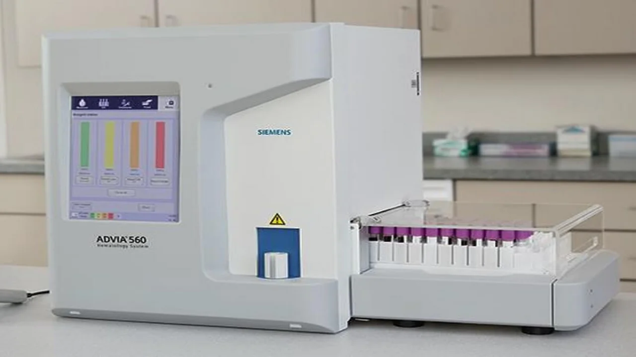
Parameters Measured by Hematology Analyzers
Explore the comprehensive guide on hematology analyzers, covering automated red cell, WBC, and platelet measurements, including key parameters and advanced features.

Hematology analyzers, particularly automated hematology analyzers, are critical tools in clinical diagnostics, offering comprehensive insights into various blood parameters. These instruments measure essential parameters such as red cell count, red cell indices (mean cell volume, mean cell hemoglobin, mean cell hemoglobin concentration), hemoglobin, hematocrit, total leukocyte count, differential leukocyte count, and platelet count. Tables 1 and 2 categorize the parameters measured by most analyzers and highlight those reported directly or through calculations. These analyzers have become an indispensable part of modern medical laboratories, helping physicians assess and diagnose numerous hematological conditions.
Most hematology analyzers measure several fundamental parameters, including RBC count, hemoglobin, mean cell volume (MCV), mean cell hemoglobin (MCH), mean cell hemoglobin concentration (MCHC), total WBC count, differential leukocyte count (either a 3-part or 5-part), and platelet count. A few analyzers provide additional measurements, such as red cell distribution width (RDW), reticulocyte count, reticulocyte hemoglobin content, mean platelet volume (MPV), platelet distribution width (PDW), and reticulated platelets.
| Parameters measured by most analyzers | Parameters measured by some analyzers |
|---|---|
|
|
Several parameters, like RBC count and hemoglobin, are either measured directly or derived from histograms. For instance, MCV and RDW are often calculated from the red cell histogram, while the leukocyte differential is obtained from a WBC histogram. Other parameters, including hematocrit, MCH, and MCHC, are derived through calculations.
| Parameters measured directly or derived through histogram | Parameters measured through calculation |
|---|---|
|
|
Hemoglobin Estimation
Hematology analyzers measure hemoglobin levels using a modified cyanmethemoglobin method, converting all hemoglobins into cyanmethemoglobin for accurate absorbance measurement at 540 nm. To reduce hazards, some analyzers employ sodium lauryl sulfate as a reagent, enabling rapid red cell lysis while minimizing interference from cell membranes and plasma lipids.
Red Blood Cell Count and Mean Cell Volume (MCV)
Red blood cells and their volume are measured using either aperture impedance or light scatter analysis. The results are displayed in a red cell histogram, where the Y-axis indicates cell number and the X-axis cell volume. The analyzer identifies red cells within the volume range of 36-360 fl. MCV aids in the morphological classification of anemia into microcytic, macrocytic, or normocytic types.

Mean Cell Hemoglobin (MCH), Mean Cell Hemoglobin Concentration (MCHC), and Hematocrit (HCT)
These key parameters are not directly measured but are calculated based on other values. The formulas used are:
- MCH (pg) = Hemoglobin (g/l) ÷ RBC count (10⁶/μl)
- MCHC (g/dl) = Hemoglobin (g/dl) ÷ Hematocrit (%)
- Hematocrit (%) = MCV (fl) ÷ RBC count (10⁶/μl)
Red Cell Distribution Width (RDW)
RDW represents the variability in red cell sizes and is expressed as a coefficient of variation of red cell size distribution. It is analogous to anisocytosis seen on a blood smear and is especially useful in distinguishing between conditions like iron deficiency anemia, where RDW is typically elevated, and β-thalassemia minor, where RDW remains normal.
Of the red cell parameters produced by the analyzer—such as red cell count, hemoglobin, hematocrit, MCV, MCH, MCHC, and RDW—the most critical for clinical decision-making are hemoglobin, hematocrit, and MCV.
WBC Differential
Difference between 3-part and 5-part hemotology analyzer...
Hematology analyzers offer either a 3-part or 5-part differential count. The 3-part differential classifies leukocytes into lymphocytes, monocytes, and granulocytes based on electrical impedance, whereas the 5-part differential provides a more detailed breakdown of leukocytes into lymphocytes, monocytes, neutrophils, eosinophils, and basophils.
Hematology analyzers utilize different methods to perform white blood cell (WBC) differentials. The 3-part differential relies on the electrical impedance volume measurement of leukocytes. In this process, the volume histogram displays cell quantities on the Y-axis and their size on the X-axis. Cells with a volume of 35-90 fl are classified as lymphocytes, those between 90-160 fl are identified as mononuclear cells, and cells with a volume ranging from 160-450 fl are labeled as neutrophils (refer to Figure 2). If the analyzer detects any irregularities in the expected histogram, it flags the result, prompting further examination via blood smear. This flagging system is designed to catch abnormalities and ensures thorough analysis, as many 3-part differential results are flagged to prevent the oversight of unusual cells.
In contrast, instruments conducting a 5-part differential operate using a combination of advanced techniques. These may include light scatter, electrical impedance, and conductivity, alongside methods such as peroxidase staining and assessing basophil resistance to lysis in acidic buffers.

Platelet Count
Platelet counting presents challenges due to their small size and tendency to aggregate. Automated hematology analyzers address this by using mathematical models to analyze platelet volume distribution. Platelet counts are typically obtained via electrical impedance, and a histogram is generated to represent the distribution, with normal platelet volume falling between 2-20 fl.
Additional platelet parameters include mean platelet volume (MPV) and platelet distribution width (PDW), which provide insights into platelet size variability. High MPV often indicates increased immature platelets in circulation, while high PDW is seen in conditions like chronic myeloid leukemia.

Hematology analyzers, including automated hematology analyzers, can provide additional platelet parameters using advanced computer technology. Two key metrics obtained from the platelet histogram are mean platelet volume (MPV) and platelet distribution width (PDW). Furthermore, some analyzers are equipped to generate an additional parameter, known as reticulated platelets.
MPV represents the average size of platelets and is derived through a mathematical calculation. The normal range for MPV is typically between 7 and 10 fl. An elevated MPV, exceeding 10 fl, indicates the presence of immature platelets in circulation. This phenomenon is often a response to peripheral platelet destruction, prompting megakaryocytes to produce larger platelets, as observed in conditions like idiopathic thrombocytopenic purpura. Conversely, a lower MPV, below 7 fl, signifies the presence of smaller platelets, which is commonly associated with reduced platelet production in the bone marrow.
PDW, analogous to red cell distribution width (RDW), measures the variation in platelet size, with normal values below 20%. An increase in PDW is typically seen in cases such as megaloblastic anemia, chronic myeloid leukemia, and post-chemotherapy patients.
Additionally, certain hematology analyzers can measure reticulated platelets, which are young platelets containing RNA, similar to reticulocytes. An increase in reticulated platelets is often observed in thrombocytopenia caused by peripheral platelet destruction, offering valuable diagnostic insights.
Reticulocyte Count
Reticulocyte counts are obtained using fluorescent dyes that bind to RNA, which is then measured by a flow cytometer. The hemoglobin content of reticulocytes is also analyzed, offering a prediction of iron deficiency.
WBC Cytogram (Scattergram)
The scattergram offers a visual representation of WBCs, with each cell type plotted according to its volume and density. This method utilizes forward angle light scatter (FALS) and side scatter (SS) to differentiate between cell types such as lymphocytes, monocytes, neutrophils, and eosinophils.
In a scattergram generated by advanced hematology analyzers, each dot symbolizes an individual cell, with its position determined by factors such as cell volume, density, side scatter, forward scatter, light absorption, and cytochemical staining, when applicable. The forward angle light scatter (FALS), representing the Y-axis, and side scatter (SS), plotted on the X-axis, provide crucial information about the cell's physical properties.
Lymphocytes, for instance, typically exhibit both low FALS and SS values. As these values increase, monocytes, neutrophils, and finally eosinophils are identified sequentially on the graph. The technology used for counting basophils, however, differs from that employed for other white blood cells, often incorporating alternative methodologies for more accurate differentiation.
The information on this page is peer reviewed by a qualified editorial review board member. Learn more about us and our editorial process.
Last reviewed on .
Article history
- Latest version
Cite this page:
- Posted by Dayyal Dungrela