Terminologies Used in Flowcytometry
Explore the key principles of flow cytometry, including fluorescence, light scatter, and data analysis, to understand how this technique identifies and analyzes cell populations.

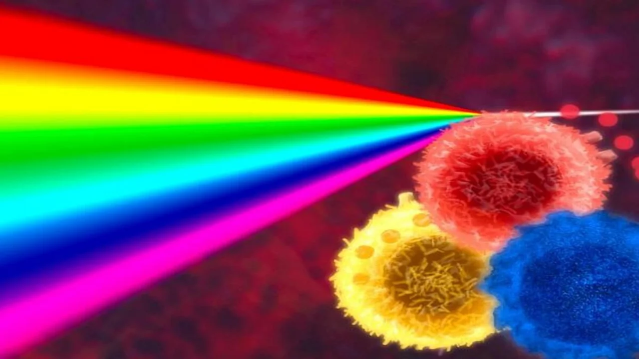
Flow cytometry is a widely used method in both research and clinical settings for analyzing various characteristics of cells or particles. By leveraging laser-based technology, it allows for the precise identification and measurement of different cell types within a sample. The technique relies on several core principles, including fluorescence, light scatter, and data analysis, which together provide a comprehensive picture of the cell populations under study.
Fluorescence
Fluorescence is a phenomenon where a fluorochrome, upon absorbing light energy, releases this energy as photon light. This characteristic allows molecules to absorb light at one wavelength and emit it at a longer wavelength. In the context of flow cytometry, commonly employed fluorescent dyes include fluorescein isothiocyanate (FITC) and phycoerythrin (PE). These fluorochrome-labeled antibodies are pivotal for detecting antigenic markers present on cell surfaces. By analyzing the antigenic profiles, specific cell types can be accurately identified. Furthermore, utilizing multiple fluorochromes enables the differentiation of various cell types within a heterogeneous cell population.
Light Scatter
When light encounters a particle within a beam, it is scattered, a process dependent on the particle's physical characteristics. In flow cytometry, two primary types of light scatter are instrumental in distinguishing cell types: forward scatter and side scatter. Forward scatter, where light continues in the same direction as the laser beam, correlates with cell size. Side scatter, where light is deflected at a 90° angle, relates to the internal complexity or granularity of the cell. These scattering patterns are crucial for identifying leukocyte subpopulations, achieved by correlating measurements of forward and side scatters. As a cell traverses the laser beam, it emits side scatter and fluorescent signals, which are captured by photomultiplier tubes, while forward scatter signals are collected by a photodiode. Both photomultiplier tubes and photodiodes function as detectors, with optical filters positioned beforehand to ensure only specific wavelengths reach them (see Figure 1).

Data Analysis
The data acquired by the flow cytometer is stored and can be presented in various formats for analysis. Parameters such as forward scatter, side scatter, or emitted fluorescence from a particle are measured as it passes through the laser beam. A histogram, one of the common display formats, plots a single parameter, with the parameter's signal value on the X-axis (horizontal axis) and the number of events on the Y-axis (vertical axis). A dot plot, on the other hand, is a two-parameter graph where each dot corresponds to a single event analyzed by the flow cytometer, with parameters displayed on both the X and Y axes. Additionally, a 3-D plot can be generated, which represents one parameter on the X-axis, another on the Y-axis, and the number of events per channel on the Z-axis.
Gating
Gating involves setting boundaries to focus the analysis on a specific population within a sample. For instance, a gate can be established on a dot plot or histogram to isolate and analyze cells that exhibit the size characteristics of lymphocytes. Gates can either be inclusive, selecting events that fall within the boundary, or exclusive, selecting events outside the boundary. The data filtered by the gate is then used in subsequent analyses.
Sorting
Typically, when a cell passes through the laser beam during flow cytometry, it is discarded as waste. However, sorting allows for the collection of specific cells of interest, defined by criteria such as size and fluorescence, for further analysis. This sorting feature is not available in all flow cytometers but is a powerful tool for applications requiring subsequent examination, such as microscopy or functional and chemical analysis.
The information on this page is peer reviewed by a qualified editorial review board member. Learn more about us and our editorial process.
Last reviewed on .
Article history
- Latest version
Cite this page:
- Posted by Dayyal Dungrela