Hematoxylin and Eosin (H&E) Staining: Principle, Procedure, and Interpretation
Step-by-step protocols for paraffin, frozen & soft tissues. Essential for cancer diagnosis and tissue analysis in histopathology.
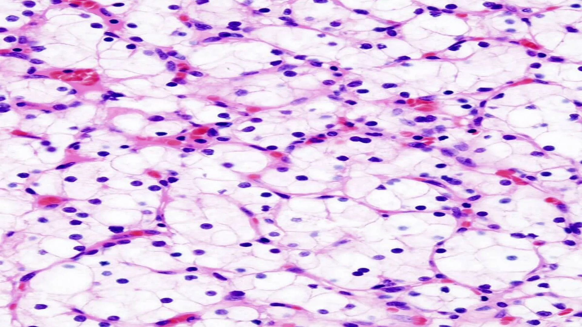
Hematoxylin and Eosin (H&E) staining is the gold standard technique in histopathology. It uses two dyes: hematoxylin (blue-purple) to stain acidic nuclear components (DNA/RNA) and eosin (pink-red) to highlight alkaline cytoplasmic proteins and extracellular structures like connective tissue. This contrast reveals tissue architecture and cellular details, enabling pathologists to diagnose diseases like cancer, assess abnormalities, and guide treatment decisions.
Hematoxylin and Eosin (H&E) Staining Principle
The principle of Hematoxylin and Eosin (H&E) staining is the differential electrostatic attraction between dyes and tissue components:
- Hematoxylin, a basic dye, binds to negatively charged (basophilic) structures like nucleic acids, staining nuclei blue-purple.
- Eosin, an acidic dye, binds to positively charged (eosinophilic) structures like proteins, staining cytoplasm and extracellular matrix pink.
This contrast reveals tissue microanatomy.
Requirements
Hematoxylin and Eosin (H&E) Staining Requirements include:
- Tissue Sections: Mounted on slides (paraffin-embedded or frozen).
- Stains:
- Hematoxylin (e.g., Harri’s: hematoxylin, ethanol, ammonium alum, mercuric oxide; or Mayer’s).
- Eosin (alcoholic eosin or formulations with eosin Y, distilled water, ethanol, and acetic acid).
- Processing Reagents:
- Deparaffinizing agents (xylene).
- Rehydration solutions (graded alcohols: 100% to 70%).
- Differentiating solution (0.5% HCl in alcohol).
- Bluing agent (ammonia water or alkaline tap water).
- Dehydration alcohols and clearing agents (xylene).
- Mounting Medium: DPX or resinous media for slide preservation.
Exact protocols vary by tissue type and laboratory standards, but these core reagents ensure high-contrast visualization of nuclei (blue) and cytoplasm/extracellular matrix (pink) for diagnostic accuracy.
Hematoxylin and Eosin (H&E) Staining Procedure
1. Procedure for Paraffin Sections (5 Micron)
Technique: Formalin-fixed, paraffin-embedded tissue sections.
- Deparaffinize & Rehydrate: Remove paraffin with xylene; hydrate through graded alcohols (100% → 70%) and water.
- Nuclear Staining: Immerse in Mayer’s hematoxylin for 1–4 minutes (time varies by tissue).
- Differentiation: Dip in 0.5% acid alcohol briefly.
- Bluing: Rinse in 1X PBS or ammonia water.
- Counterstain: Apply alcoholic-eosin (1% Eosin Y + Phloxine B) for 30 seconds–1 minute.
- Dehydration & Mounting: Dehydrate through alcohols, clear in xylene, and mount with DPX.
Note: Do NOT rinse with water after eosin! Proceed directly to dehydration.
2. Procedure for Fresh Frozen Sections
Technique: Unfixed frozen sections requiring immediate fixation.
- Fixation: Place slides in cold 10% neutral buffered formalin or 4% PFA for 10 minutes.
- Remove OCT Compound: Rinse in 1X PBS and tap water.
- Nuclear Staining: Mayer’s hematoxylin for 30 seconds.
- Bluing: 1X PBS for 20 seconds.
- Counterstain: Alcoholic-eosin for 30 seconds.
- Dehydration & Mounting: Dehydrate through alcohols, clear in xylene, and mount with DPX.
Difference: Includes fixation to preserve fresh tissue.
3. Procedure for Frozen Sections (Non-Fresh)
Technique: Pre-fixed frozen sections.
- Rinse OCT: Wash slides in 1X PBS for 3 minutes to remove OCT compound.
- Nuclear Staining: Mayer’s hematoxylin for 30 seconds.
- Bluing: 1X PBS for 20 seconds.
- Counterstain: Alcoholic-eosin for 30 seconds.
- Dehydration & Mounting: Dehydrate through alcohols, clear in xylene, and mount with DPX.
Difference: Skips fixation step.
4. Procedure for Soft Tissue Paraffin Sections (4 Micron)
Technique: Formalin-fixed, paraffin-embedded soft tissue sections.
- Deparaffinize & Rehydrate: Remove paraffin with xylene; hydrate through graded alcohols (100% → 70%) and water.
- Extended Nuclear Staining: Hematoxylin for 4 minutes (longer for dense soft tissues).
- Differentiation: 0.5% acid alcohol (1–2 dips).
- Counterstain: Eosin for 30 seconds.
- Mounting: Dehydrate through alcohols, clear in xylene, and mount with DPX.
Difference: Extended hematoxylin incubation for better nuclear penetration in fibrous tissues.
Why All Four Procedures Matter
- Paraffin-Embedded Tissues: Standard method for preserved tissues (e.g., biopsies).
- Fresh Frozen Sections: For rapid diagnosis (e.g., intraoperative consultations).
- Frozen Sections: For pre-fixed frozen tissues (e.g., research samples).
- Soft Tissue: Optimized for dense tissues (e.g., muscle, cartilage).
Result and Interpretation
Hematoxylin and Eosin (H&E) staining produces a high-contrast visualization of tissue architecture, enabling pathologists to analyze cellular and extracellular components. The results are consistent across all sample preparation methods:
- Nuclei: Blue (due to hematoxylin).
- Cytoplasm: Pink (due to eosin).
- Erythrocytes: Bright red.
- Other eosinophilic structures: Red, pink, or orange.
Key Observations
- Nuclear Details (Blue/Purple):
- Normal: Uniform nuclei with fine chromatin (healthy cells).
- Abnormal: Enlarged, irregular nuclei; hyperchromasia (dark staining); prominent nucleoli (e.g., cancer cells).
- Cytoplasmic & Extracellular Components (Pink/Red):
- Normal: Smooth cytoplasmic boundaries; intact collagen/connective tissue.
- Abnormal: Cytoplasmic vacuolation (e.g., fatty liver), disrupted collagen (fibrosis), or necrosis (loss of cellular detail).
- Diagnostic Insights:
- Cancer: Nuclear atypia, high mitotic activity, irregular glandular structures.
- Inflammation: Neutrophils, lymphocytes, or plasma cells infiltrating tissue.
- Infections: Granulomas (e.g., tuberculosis), viral inclusions (e.g., CMV).
Observations
- Tissue Integrity: Preservation of architecture (normal) vs. disorganization (disease).
- Artifacts: Over-staining, folding, or shrinkage (indicate technical errors).
Diagnostic Significance
The interpretation of H&E-stained slides enables the identification of a wide range of abnormalities, including inflammation, neoplasia, and degenerative changes. For example, hyperchromatic and pleomorphic nuclei with disrupted cell arrangement might indicate cancer, while increased extracellular matrix staining could suggest fibrosis.
FAQs
What are hematoxylin and eosin?
How does hematoxylin and eosin staining work?
What do hematoxylin and eosin stain?
How to prepare hematoxylin and eosin staining?
What is hematoxylin and eosin staining used for?
Why is hematoxylin and eosin staining used?
The information on this page is peer reviewed by a qualified editorial review board member. Learn more about us and our editorial process.
Last reviewed on .
Article history
- Latest version
Cite this page:
- Posted by Dayyal Dungrela