Myeloperoxidase (MPO) Stain: Purpose, Principle, Procedure, and Interpretation
Discover the MPO stain, essential for acute myeloid leukemia diagnosis. Learn its purpose, procedure, and how it differentiates myeloid from lymphoid cells.
Myeloperoxidase (MPO) is a lysosomal enzyme found in the azurophilic granules of neutrophils, eosinophils, and some monocytes. MPO generates reactive oxygen species to destroy pathogens and enhance the immune response. Lymphocytes do not contain MPO.
Myeloperoxidase (MPO) stain is a cytochemical test used in hematology to detect MPO enzyme in neutrophils, monocytes, and myeloid precursors. It is the gold standard for differentiating acute myeloid leukemia (AML) from acute lymphoblastic leukemia (ALL) and diagnosing MPO deficiency in neutrophils.
Purpose of Myeloperoxidase Stain
The primary purpose of the MPO stain is to distinguish between myeloid and lymphoid cell lineages. This distinction is essential in the diagnosis and classification of leukemias.
- Differentiation of Blood Cell Types: MPO staining helps identify myeloid cells by indicating the presence of the myeloperoxidase enzyme, which is absent in lymphoid cells.
- Diagnosis of Acute Myeloid Leukemia (AML): In AML, a significant proportion of blasts (immature cells) exhibit MPO positivity, whereas in acute lymphoblastic leukemia (ALL), the blasts are typically MPO negative. This difference assists in distinguishing between AML and ALL.
- Assessment of Myeloperoxidase Deficiency: MPO staining can be used to diagnose congenital myeloperoxidase deficiency, a condition characterized by a lack of MPO enzyme in neutrophils.
Principle of Myeloperoxidase (MPO) Stain
The principle of the Myeloperoxidase (MPO) stain is based on the peroxidase activity of the myeloperoxidase enzyme. In the presence of hydrogen peroxide, MPO catalyzes the oxidation of benzidine derivatives (e.g., 3,3′-diaminobenzidine), leading to the formation of a blue or brown colored derivative that marks MPO-positive cells.
- Chemical Reaction: MPO oxidizes substrates such as benzidine or 3,3′-diaminobenzidine in the presence of hydrogen peroxide, resulting in an insoluble blue or brown derivative.
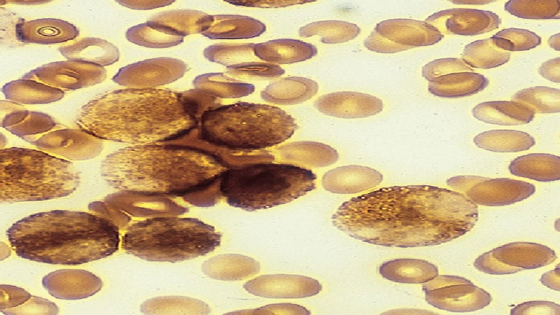
- Staining Pattern: Cells containing MPO exhibit a positive staining reaction, appearing dark brown or blue-black under the microscope, while MPO-negative cells remain unstained.
Myeloperoxidase Stain Procedure
Materials and Reagents Required
- Air-dried blood/bone marrow smears.
- Fixative (e.g., formalin)
- MPO staining kit (benzidine, H₂O₂, counterstain).
- Buffer solution
Myeloperoxidase Staining Protocol
- Fixation: Use 10% formalin for 30 seconds.
- Incubation: Apply benzidine-H₂O₂ solution (10–15 minutes).
- Counterstain: Use hematoxylin to provide contrast for cellular components.
- Microscopy: Assess for brown cytoplasmic granules.
Interpretation of Myeloperoxidase Stain Results
MPO Stain Positive vs. Negative
- Positive: Brown granular deposits (≥3% blasts in AML).
- Negative: No staining (suggests lymphoid origin).
Grading System for MPO Activity
| Grade | Description | Clinical Relevance |
|---|---|---|
| 0 | No granules | Lymphoid origin |
| 1+ | Faint granules | Early myeloid differentiation |
| 2+ | Moderate positivity | Typical AML |
| 3+ | Strong positivity | High enzymatic activity |
Role of MPO Stain in Diagnosing Leukemia
MPO Stain in Acute Myeloid Leukemia (AML): Over 80% of AML cases show myeloperoxidase stain-positive blasts, confirming myeloid lineage. MPO-negative AML is rare but linked to specific genetic mutations.
MPO-Negative Leukemias: MPO negativity aids in excluding AML, directing focus to ALL or mixed-phenotype acute leukemia (MPAL).
MPO Stain in Bone Marrow Analysis
Evaluating Myeloid Lineage Cells: In mpo stain bone marrow assessments, MPO positivity in >3% blasts confirms AML. Correlate with flow cytometry for accuracy.
MPO Staining in Neutrophils: Clinical Relevance
Detecting Neutrophil Dysfunction: Abnormal mpo staining neutrophils indicate chronic granulomatous disease or myelodysplastic syndromes.
Common Challenges in MPO Staining
Addressing False Results
- False Negatives: Over-fixation or inactive reagents.
- False Positives: Contamination or endogenous peroxidases (e.g., erythrocytes).
FAQs
How long does MPO staining take?
Can MPO stain diagnose non-hematological disorders?
What are MPO stain limitations?
The information on this page is peer reviewed by a qualified editorial review board member. Learn more about us and our editorial process.
Last reviewed on .
Article history
- Latest version
Cite this page:
- Posted by Dayyal Dungrela