Microscopic Examination of Urine
Learn about the various cells and components found in human urine, including WBCs, RBCs, different types of epithelial cells, casts, crystals, bacteria, and more.

The microscopic analysis of urine is often referred to as the “liquid biopsy of the urinary tract.”
Urine comprises diverse microscopic, insoluble, solid components in suspension. These components fall into two categories: organized and unorganized. Organized substances encompass red blood cells, white blood cells, epithelial cells, casts, bacteria, and parasites. On the other hand, unorganized substances consist of crystalline and amorphous material. These elements remain suspended in urine, settling down and forming sediment at the container's bottom over time. Consequently, they are termed urinary deposits or urinary sediments. The examination of these deposits proves valuable in diagnosing urinary tract diseases, as outlined in Table 1.
| Condition | Albumin | RBCs/HPF | WBCs/HPF | Casts/LPF | Others |
|---|---|---|---|---|---|
| Normal | 0-trace | 0-2 | 0-2 | Occasional (Hyaline) | – |
| Acute glomerulonephritis | 1-2+ | Numerous; dysmorphic | 0-few | Red cell, granular | Smoky urine or hematuria |
| Nephrotic syndrome | > 4+ | 0-few | 0-few | Fatty, hyaline, Waxy, epithelial | Oval fat bodies, lipiduria |
| Acute pyelonephritis | 0-1+ | 0-few | Numerous | WBC, granular | WBC clumps, bacteria, nitrite test |
| HPF: High power field; LPF: Low power field; RBCs: Red blood cells; WBCs: White blood cells. | |||||
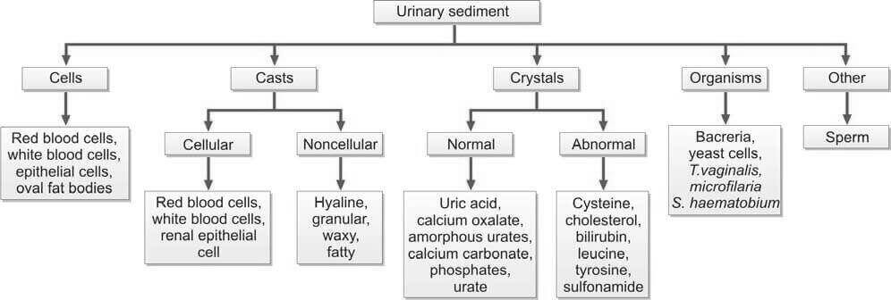
Various categories of urinary sediments are illustrated in Figure 1. The primary objective of the microscopic analysis of urine is to discern distinct types of cellular elements and casts. It's noteworthy that the majority of crystals observed have minimal clinical significance.
Specimen
Cellular elements exhibit optimal preservation in acid, hypertonic urine, while they degrade swiftly in an alkaline, hypotonic solution. A mid-stream, freshly voided, first-morning specimen is the preferred choice due to its heightened concentration. It is crucial to examine the specimen within 2 hours of voiding, as cells and casts undergo degeneration when left standing at room temperature. If a preservative is necessary, the addition of 1 crystal of thymol or 1 drop of formalin (40%) to approximately 10 ml of urine is recommended.
Method
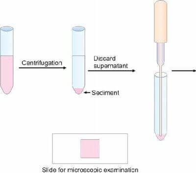
A thoroughly mixed urine sample (12 ml) undergoes centrifugation in a centrifuge tube for 5 minutes at 1500 rpm, and the supernatant is carefully poured off. To resuspend the sediment, the tube is tapped at the bottom, utilizing 0.5 ml of urine. A small droplet of this resuspended sediment is placed on a glass slide and covered with a cover slip, as depicted in Figure 2. Subsequently, the slide is promptly examined under the microscope, initially using the low-power objective and then transitioning to the high-power objective. For enhanced visualization of the elements, it is advisable to lower the condenser, thereby reducing the illumination.
Cells
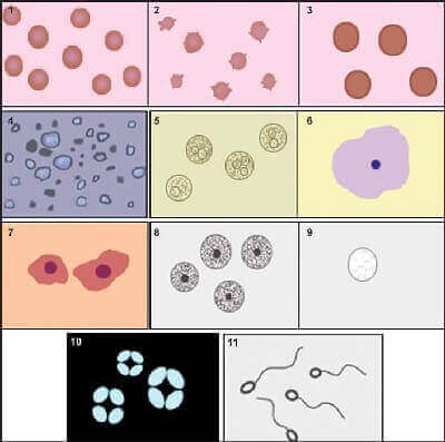
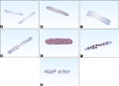
Figure 3 illustrates the cellular elements present in urine.
Red Blood Cells
Normally, urine contains either no red blood cells or only an occasional one. In a fresh urine sample, red cells manifest as small, smooth, yellowish, anucleate biconcave disks measuring approximately 7 μ in diameter, commonly referred to as isomorphic red cells. However, in dilute or hypotonic urine, these red cells may appear swollen, taking on the form of thin discs with a larger diameter (9-10 μ). Conversely, in hypertonic urine, they may exhibit a crenated appearance, characterized by a smaller diameter and a spiky surface.
In cases of glomerulonephritis, red cells are often described as dysmorphic, indicating a marked variability in size and shape. This distortion arises from the passage of red cells through damaged glomeruli. The presence of over 80% dysmorphic red cells strongly suggests glomerular pathology.
The quantification of red cells can be reported as the number of red cells per high-power field. The causes of hematuria have been previously outlined.
White Blood Cells (Pus Cells)
White blood cells typically exhibit a spherical form, measuring 10-15 μ in size, and display a granular appearance where nuclei may be discernible. Degenerated white cells, on the other hand, appear distorted, smaller, and possess fewer granules. In instances of infection, clumps of numerous white cells become evident. The presence of a substantial number of white cells in urine is termed pyuria. In hypotonic urine, white cells swell, and their granules become highly refractile, exhibiting Brownian movement. These cells, referred to as glitter cells, indicate injury to the urinary tract, particularly when present in large numbers.
Under normal conditions, 0-2 white cells may be observed per high-power field. However, the detection of pus cells exceeding 10 per high-power field or the presence of clumps suggests a potential urinary tract infection.
Elevated white cell counts are associated with conditions such as fever, pyelonephritis, lower urinary tract infection, tubulointerstitial nephritis, and renal transplant rejection.
In cases of urinary tract infection, a combination of the following is typically observed:
- Clumps of pus cells or pus cells exceeding 10 per high-power field
- Presence of bacteria
- Albuminuria
- Positive nitrite test
The simultaneous presence of white cells and white cell casts is indicative of renal infection, specifically pyelonephritis.
Eosinophils, constituting more than 1% of urinary leucocytes, serve as a characteristic feature of acute interstitial nephritis due to a drug reaction, a detail better appreciated with Wright's stain.
Renal Tubular Epithelial Cells
The identification of renal tubular epithelial cells holds significant diagnostic value. Elevated counts of these cells are indicative of conditions causing tubular damage, such as acute tubular necrosis, pyelonephritis, viral kidney infections, allograft rejection, and poisoning from substances like salicylate or heavy metals.
These cells exhibit characteristics of being small, approximately the same size or slightly larger than white blood cells. They can take on a polyhedral, columnar, or oval shape, featuring granular cytoplasm. Often, a single, large, refractile, eccentric nucleus is observable.
Distinguishing renal tubular epithelial cells from pus cells in unstained preparations can pose a challenge due to their similarity in appearance. This makes the identification process in such conditions a meticulous task requiring close examination.
Squamous Epithelial Cells
Squamous epithelial cells form the lining of the lower urethra and vagina. Optimal visibility is achieved under a low-power objective (×10). When a substantial quantity of squamous cells is detected in urine, it suggests contamination of the urine with vaginal fluid. These cells are characterized by their size, being large and rectangular, with a flat morphology, abundant cytoplasm, and a small, central nucleus.
Transitional Epithelial Cells
Transitional cells form the lining of the renal pelvis, ureters, urinary bladder, and upper urethra. These cells, often referred to as caudate cells due to their diamond- or pear-shaped appearance, are notably large. The presence of abundant transitional cells or the observation of cell sheets in urine is commonly associated with scenarios such as catheterization procedures and cases of transitional cell carcinoma.
Oval Fat Bodies
These represent degenerated renal tubular epithelial cells laden with highly refractile lipid, primarily composed of cholesterol droplets. When examined under polarized light, these cells exhibit a distinctive “Maltese cross” pattern. To facilitate identification, they can be stained using a fat stain like Sudan III or Oil Red O. This phenomenon is commonly observed in nephrotic syndrome, a condition characterized by the presence of lipiduria.
Spermatozoa
Occasionally, these elements may be observed in the urine of males.
Telescoped Urinary Sediment: This term denotes a urinary sediment composition comprising red blood cells, white blood cells, oval fat bodies, and various types of casts in approximately equal proportions. This phenomenon is associated with conditions such as lupus nephritis, malignant hypertension, rapidly proliferative glomerulonephritis, and diabetic glomerulosclerosis.
Organisms
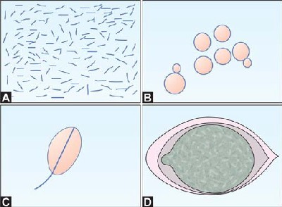
The organisms identifiable in urine are depicted in Figure 4.
Bacteria
Detection of bacteria in urine can be accomplished through microscopic examination, reagent strip tests assessing for significant bacteriuria (utilizing the nitrite test and leucocyte esterase test), as well as culture.
Significant bacteriuria is confirmed when the bacterial colony-forming units/ml of urine surpass specific thresholds:
- In a clean-catch midstream sample, it is present when there are more than 10^5 bacterial colony-forming units per milliliter (cfu/ml) of urine.
- In a catheterized sample, the threshold is set at over 10^4 cfu/ml of urine.
- For a suprapubic aspiration sample, significant bacteriuria is considered when there are more than 10^3 cfu/ml of urine.
Tests for the Detection of Bacteria in Urine
- Microscopic Examination: In a wet preparation, the documentation of bacteria presence is pertinent only when the urine is freshly collected. Bacteria are typically observed in conjunction with pus cells. A Gram's-stained smear of uncentrifuged urine revealing one or more bacteria per oil-immersion field suggests the likelihood of over 10^5 bacterial colony-forming units per milliliter (cfu/ml) of urine. If numerous squamous cells are present, there's a probability of urine contamination with vaginal flora. Additionally, the sole presence of bacteria without pus cells indicates potential contamination with vaginal or skin flora.
- Chemical or Reagent Strip Tests for Significant Bacteriuria: These tests utilize strips saturated with specialized chemical reagents that interact with components linked to bacterial existence in urine. These strips commonly evaluate the presence of nitrites, signaling bacterial metabolism, and leukocyte esterase, an enzyme released by white blood cells in response to infection.
- Culture: Upon culturing, a colony count exceeding 10^5/ml strongly indicates a urinary tract infection, even in asymptomatic females. A positive culture is subsequently followed by sensitivity testing. The majority of infections are attributed to Gram-negative enteric bacteria, notably Escherichia coli.
Identification of three or more bacterial species in a culture typically suggests contamination by vaginal flora.
A lack of bacterial growth in the presence of pyuria, often referred to as ‘sterile’ pyuria, is associated with various conditions. These include prior antibiotic therapy, renal tuberculosis, prostatitis, renal calculi, catheterization, fever in children (regardless of the underlying cause), female genital tract infection, and non-specific urethritis in males.
Yeast Cells (Candida)
These structures exhibit a round or oval morphology, closely mirroring the size of red blood cells. Unlike red cells, they display a distinctive budding pattern, possess an oval and highly refractile appearance, and are insoluble in 2% acetic acid.
The detection of Candida in urine may indicate an immunocompromised condition, the presence of vaginal candidiasis, or diabetes mellitus. Typically, the presence of pyuria accompanies Candida infection. However, it's crucial to note that Candida could also be a potential contaminant in the sample. Therefore, the urine sample must be examined in a fresh state to ensure accurate interpretation.
Trichomonas vaginalis
These microorganisms exhibit motility and possess a distinctive pear shape, featuring an undulating membrane on one side and four flagellae. Their presence is associated with vaginitis in females, making them contaminants in urine samples. Notably, their motility renders them easily detectable in freshly collected urine.
Eggs of Schistosoma haematobium
The presence of Schistosoma haematobium eggs is notably widespread in Egypt. This parasitic organism, responsible for causing schistosomiasis, particularly affects the urinary tract. The eggs of Schistosoma haematobium are commonly found in human urine, indicating an established infection.
Schistosomiasis, also known as bilharzia, is a waterborne disease prevalent in regions with inadequate sanitation and limited access to clean water. In Egypt, where conditions conducive to the transmission of this parasitic infection exist, the prevalence underscores the public health challenges associated with waterborne diseases in the region.
Effective control measures, including improved water sanitation and targeted medical interventions, are crucial to mitigating the impact of Schistosoma haematobium infections in affected populations.
Microfilariae
Microfilariae, minute larvae of filarial parasites, may be observed in urine, particularly in cases of chyluria. Chyluria arises from the rupture of urogenital lymphatic vessels, allowing the infiltration of chyle, a milky fluid rich in lymphocytes and lipids, into the urinary system.
The presence of microfilariae in urine samples is a diagnostic marker for certain filarial infections, with different species exhibiting distinct characteristics. Common culprits include Wuchereria bancrofti and Brugia malayi, both known to cause lymphatic filariasis in affected individuals.
Clinical assessment and laboratory analysis of urine containing microfilariae play a crucial role in confirming the diagnosis and guiding appropriate treatment strategies. Management often involves antiparasitic medications and supportive care to address the underlying lymphatic pathology.
Casts
Urinary casts represent cylindrical, cigar-shaped microscopic structures originating within the distal renal tubules and collecting ducts. These formations mirror the shape and diameter of the lumina, essentially serving as molds or ‘casts’ of the renal tubules. Exhibiting parallel sides and rounded ends, the dimensions of these casts can vary. Composed primarily of a precipitate of Tamm-Horsfall protein, a protein secreted by tubules, their formation is confined to the renal tubules. Consequently, the presence of casts serves as an indicative marker of renal parenchymal disease.
While various types of casts exist, all urine casts fundamentally possess a hyaline composition. The diverse types emerge as different elements deposit onto the hyaline material, as illustrated in Figure 5. Optimal visualization of casts is achieved under a low-power objective (×10), with the condenser lowered to diminish illumination.

Casts constitute the exclusive components within the urinary sediment that distinctly originate from the renal system.
There are two primary categories of casts (refer to Figure 6):
- Noncellular: This includes hyaline, granular, waxy, and fatty casts.
- Cellular: Encompassing red blood cell, white blood cell, and renal tubular epithelial cell casts.
Hyaline and granular casts may manifest in both normal and pathological conditions. In contrast, casts featuring red blood cells, white blood cells, or renal tubular epithelial cells are indicative of kidney diseases.
Non-cellular Casts
Hyaline Casts: This type of cast is the most prevalent in urine, characterized by a homogenous, colorless, transparent, and refractile appearance. Hyaline casts present as cylindrical structures with parallel sides, blunt rounded ends, and a low refractive index. Occasional hyaline casts are considered normal, but an increased presence, known as “cylinduria”, is indicative of abnormal conditions. Composed primarily of Tamm-Horsfall protein, they may transiently appear after strenuous exercise or during fever. Elevated numbers are associated with conditions causing glomerular proteinuria.
Granular Casts: The presence of degenerated cellular debris imparts a granular appearance to these cylindrical structures. Featuring coarse or fine granules representing degenerated renal tubular epithelial cells embedded in a Tamm-Horsfall protein matrix, granular casts are observed after strenuous exercise, in cases of fever, acute glomerulonephritis, and pyelonephritis.
Waxy Casts: Easily recognizable, waxy casts form when hyaline casts linger in renal tubules for an extended period (prolonged stasis). Exhibiting a homogenous, smooth, glassy appearance with cracked or serrated margins and irregular, broken-off ends, waxy casts are distinct. Unlike other casts, their ends are straight and sharp. Typically light yellow in color, waxy casts are frequently observed in end-stage renal failure.
Fatty Casts: Comprising cylindrical structures filled with highly refractile fat globules, mainly triglycerides and cholesterol esters within a Tamm-Horsfall protein matrix, fatty casts are characteristic of nephrotic syndrome.
Broad Casts: These casts form in dilated distal tubules and are associated with chronic renal failure and severe renal tubular obstruction. Both waxy and broad casts are linked to a poor prognosis.
Cellular Casts
To be classified as cellular, casts must contain a minimum of three cells within the matrix. The nomenclature of cellular casts is contingent upon the specific type of cells entrapped in the matrix.
Red Cell Casts: These cylindrical structures exhibit red blood cells embedded in a Tamm-Horsfall protein matrix. Their coloration may range to brown due to hemoglobin pigmentation. Red cell casts hold paramount diagnostic significance, particularly in distinguishing hematuria stemming from glomerular disease as opposed to other causes. The presence of RBC casts typically indicates glomerular pathology, such as acute glomerulonephritis.
White Cell Casts: These cylindrical formations feature white blood cells encased in a Tamm-Horsfall protein matrix. Leukocytes generally traverse into tubules from the interstitium; thus, the presence of white cell casts is indicative of tubulointerstitial disease, such as pyelonephritis.
Renal Tubular Epithelial Cell Casts: Comprising sloughed-off renal tubular epithelial cells, these casts are observed in conditions like acute tubular necrosis, viral renal disease, heavy metal poisoning, and acute allograft rejection. Even the occasional detection of renal tubular casts holds significant diagnostic value.
Crystals
Crystals represent refractile structures characterized by a distinct geometric shape, resulting from the orderly three-dimensional arrangement of atoms and molecules. In contrast, amorphous material lacks a definite shape and typically presents as granular aggregates or clumps.
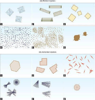
Urine crystals, as illustrated in Figure 7, can be classified into two primary types: (1) Normal, observed in the urinary sediment of healthy individuals, and (2) Abnormal, prevalent in states of disease.
Nevertheless, crystals identified in normal urine may manifest in elevated quantities in certain diseases.
While the majority of crystals, especially phosphates, urates, and oxalates, generally lack clinical significance, their increased presence can be indicative of underlying conditions. The identification of crystals in urine relies on their distinctive morphology. However, before confirming the existence of any abnormal crystals, it is imperative to validate their presence through chemical tests.
Normal Crystals
Crystals found in acidic urine:
- Uric Acid Crystals: Displaying variable shapes such as diamond, rosette, and plates, these crystals exhibit a yellow or red-brown coloration attributed to urinary pigments. Soluble in alkali but insoluble in acid, increased numbers are associated with conditions like gout and leukemia. It's noteworthy that flat hexagonal uric acid crystals may be mistaken for cysteine crystals, which also form in acidic urine.
- Calcium Oxalate Crystals: Manifesting as colorless, refractile, and envelope-shaped structures, these crystals may also assume dumbbell-shaped or peanut-like forms. Soluble in dilute hydrochloric acid, an elevation in their numbers, known as oxaluria, may suggest the presence of oxalate stones. Certain food intake, such as tomatoes, spinach, cabbage, asparagus, and rhubarb, can contribute to increased crystal numbers. Moreover, a substantial quantity is observed in cases of ethylene glycol poisoning.
- Amorphous Urates: Comprising urate salts of potassium, magnesium, or calcium in acidic urine, these crystals typically appear as yellow, fine granules in compact masses. Their solubility in alkali or saline at 60°C distinguishes them within the crystalline spectrum.
Crystals found in alkaline urine:
- Calcium Carbonate Crystals: Small, colorless, and often found in pairs, these crystals demonstrate solubility in acetic acid, releasing gas bubbles upon dissolution.
- Phosphates: Phosphates may manifest as crystals, such as triple phosphates and calcium hydrogen phosphate, or as amorphous deposits.
- Triple Phosphates (Ammonium Magnesium Phosphate): Colorless and exhibiting a shiny, 3-6 sided prismatic structure with oblique surfaces at the ends, often resembling “coffin lids”, or presenting a feathery fern-like appearance.
- Calcium Hydrogen Phosphate (Stellar Phosphate): Colorless and displaying variable shapes, such as star-shaped formations, plates, or prisms.
- Amorphous Phosphates: Occurring as colorless, small granules, often dispersed. All phosphates exhibit solubility in dilute acetic acid.
- Ammonium Urate Crystals: Presenting as cactus-like structures covered with spines, these crystals, often referred to as ‘thornapple’ crystals, are yellow-brown and soluble in acetic acid at 60°C.
Abnormal Crystals
Although rare, the presence of these crystals indicates an underlying pathological process.
These crystals typically emerge in an acidic pH, often in substantial quantities. However, reporting abnormal crystals based solely on microscopy is insufficient; additional chemical tests are essential for confirmation.
- Cysteine Crystals: Colorless, transparent, and hexagonal with six sides, these highly refractile plates are commonly found in acid urine. Frequently occurring in layered formations, they dissolve in 30% hydrochloric acid. Cysteine crystals are associated with cystinuria, an inborn metabolic error, and are often linked to the formation of cysteine stones.
- Cholesterol Crystals: Colorless and refractile, these flat rectangular plates feature notched corners, arranged in a stair-step pattern. Soluble in ether, chloroform, or alcohol, they are observed in lipiduria, such as nephrotic syndrome and hypercholesterolemia. Identification can be confirmed with a polarizing microscope.
- Bilirubin Crystals: Small (5 μ) and brown, these crystals exhibit variable shapes, including square, bead-like, or fine needles. Confirmation of their presence is achieved through reagent strip or chemical tests for bilirubin. Soluble in strong acid or alkali, they are indicative of severe obstructive liver disease.
- Leucine Crystals: Refractile and yellow or brown, these crystals manifest as spheres with radial or concentric striations. Soluble in alkali, they are commonly found in urine alongside tyrosine in cases of severe liver disease, such as cirrhosis.
- Tyrosine Crystals: Appearing as clusters of fine, delicate, colorless, or yellow needles, these crystals are observed in liver disease and tyrosinemia, an inborn metabolic error. They dissolve in alkali.
- Sulfonamide Crystals: Exhibiting variable shapes but often appearing as sheaves of needles, these crystals arise following sulfonamide therapy. They are soluble in acetone.
The information on this page is peer reviewed by a qualified editorial review board member. Learn more about us and our editorial process.
Last reviewed on .
Article history
- Latest version
Cite this page:
- Comment
- Posted by Dayyal Dungrela