Sudan Black B Stain: Purpose, Principle, Procedure, and Interpretation
Learn about Sudan Black B Stain—its purpose, principle, procedure, and interpretation. Discover how this essential stain aids in diagnosing leukemia and other hematological disorders.
Sudan Black B (SBB) is a lipid-soluble dye used in hematology and histology to identify lipid-containing granules in granulocytes, monocytes, and other cells. It helps distinguish myeloid from lymphoid cells in leukemia diagnostics, offering results comparable to myeloperoxidase (MPO) staining. Its ability to bind irreversibly to lipids provides important details about cell composition, making it essential for studying hematological disorders.
Sudan Black B Stain Composition
Sudan Black B stain contains Sudan Black B dye, a fat-soluble stain, dissolved in 70% ethanol or propylene glycol. It selectively stains lipids in cells and tissues. Some protocols may include a counterstain like hematoxylin to enhance contrast.
Purpose of Sudan Black B Stain
Sudan Black B stain is commonly used for:
- Diagnosing hematological malignancies such as acute myeloid leukemia (AML).
- Differentiating between myeloid and lymphoid lineages in blood and bone marrow smears.
- Detecting lipid granules and substances in various cells and tissues.
Advantages of Sudan Black B (SBB) Stain Over MPO Stain
- Enhanced Lipid Detection: Sudan Black B cytochemical stain differentiates acute myeloid leukemia (AML) from acute lymphoid leukemia (ALL) by selectively staining lipids in myeloid cell granules. AML cells stain intensely due to their higher lipid content, while ALL cells show little to no staining, aiding in leukemia diagnosis.
- Resistance to Fixation Artifacts: SBB is less sensitive to fixation artifacts, ensuring greater accuracy and reliability compared to MPO.
- Stain Longevity: SBB stains show minimal fading over time and can be used on smears over two weeks old.
- Granule Staining: SBB stains both azurophilic and specific granules in neutrophils, unlike MPO, which stains only azurophilic granules.
Principle of Sudan Black B Stain
Sudan Black B (SBB) is a lipid-soluble dye that stains lipids like sterols, neutral fats, and phospholipids found in azurophilic granules of myeloid cells and lysosomes in monocytic cells. The dye binds to these lipids due to its higher solubility in lipids than in the solvent, resulting in black pigmentation under microscopy when the stain is absorbed by lipid-rich cellular components.
Important points about the Sudan Black B stain principle include:
- It is a fat-soluble dye that penetrates the lipid membranes of cells, staining them darkly.
- Lipid-containing granules in myeloid cells stain more prominently, making it easier to differentiate these cells from lymphoid cells.
- Compared to other stains like myeloperoxidase (MPO), Sudan Black B is less sensitive to fixation artifacts, making it a preferred choice in many labs.
Reagents and Materials Required
For the Sudan Black B stain procedure, the following reagents and materials are essential:
- Sudan Black B dye (prepared in ethanol or isopropanol).
- Buffer solutions for pH stabilization.
- Microscope slides and coverslips.
- Fixatives (e.g., methanol or formalin) to preserve the specimen.
- Counterstain (May–Grünwald–Giemsa or Leishman stain).
- Specimen samples (blood smears or bone marrow aspirates).
Proper storage of reagents and adherence to preparation protocols are crucial for obtaining accurate results.
Procedure of Sudan Black B Stain
- Fixation: Fix air-dried smears in formalin vapor, formaldehyde, or formalin-ethanol for 10 minutes.
- Washing: Rinse gently in water for 5–10 minutes.
- Staining: Immerse slides in Sudan Black B solution for 1 hour in a covered Coplin jar.
- Differentiation: Rinse with 70% alcohol for 30 seconds, repeating three times.
- Rinsing & Drying: Wash in running tap water and air dry.
- Counterstaining: Apply Leishman stain or May–Grünwald–Giemsa stain without further fixation.
- Microscopic Examination: Air dry completely and examine under a microscope.
Interpretation of Sudan Black B Stain Results
Positive Results
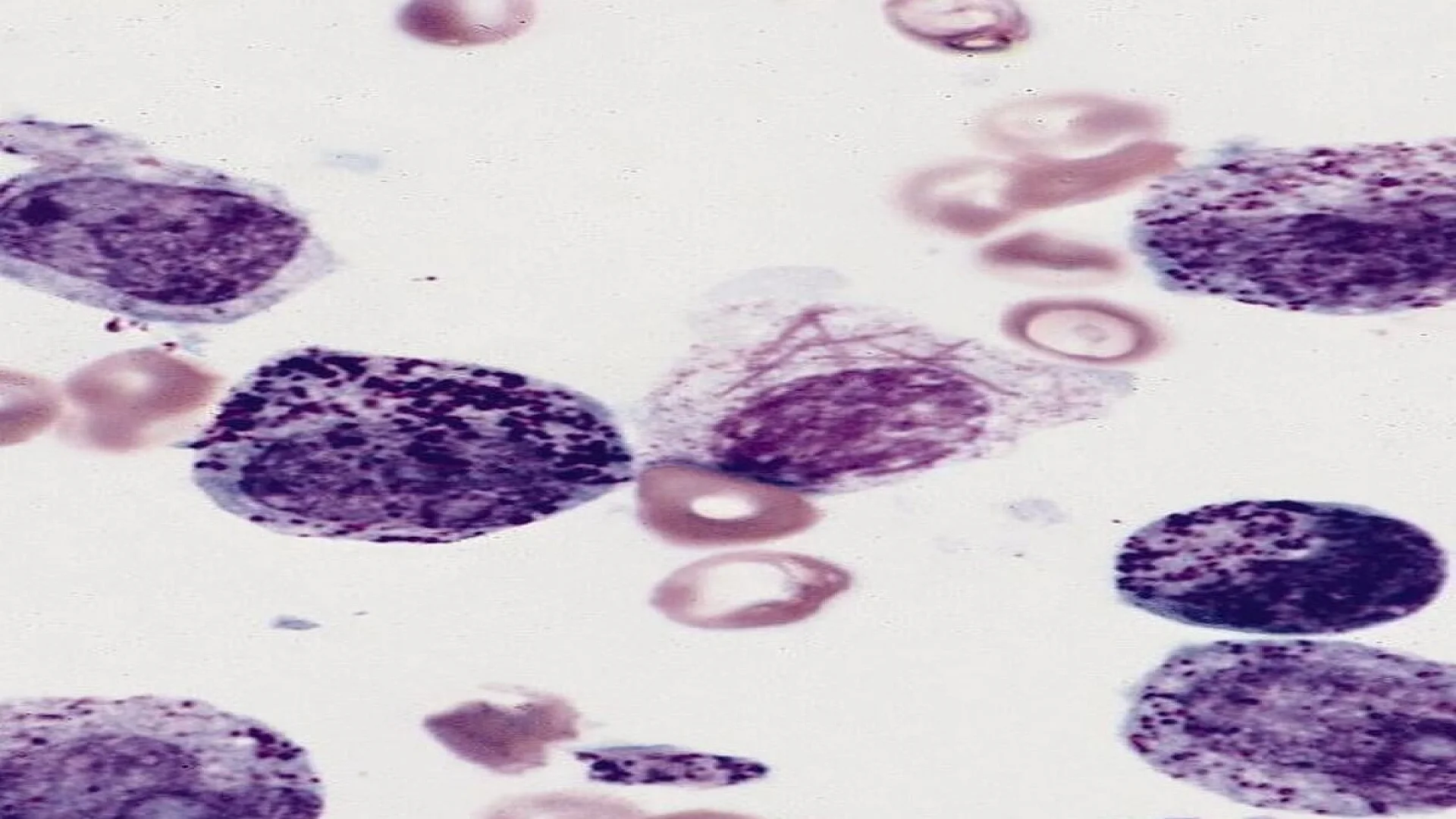
- Cells containing lipid-rich granules (e.g., myeloid cells) appear dark blue to black.
- Sudan Black B stain-positive findings are typically associated with myeloid malignancies, including AML.
Negative Results
- Lymphoid cells and other non-lipid-containing cells do not retain the stain, appearing light or unstained.
- Negative results may prompt additional testing to confirm the diagnosis.
Limitations of Sudan Black B Stain
Sudan Black B (SBB) has limited specificity, requiring expert interpretation to avoid false positives. It is not suitable for paraffin-embedded tissues and may cause non-specific staining due to its fat-soluble nature. SBB does not stain non-lipid-containing cells and requires careful handling due to its potential to stain skin and clothing.
FAQs
What is the principle of Sudan Black B stain?
How is Sudan Black B stain used for leukemia?
What are the steps in Sudan Black B stain procedure?
In which type of leukemia is the Sudan Black B stain positive?
What substance in the blood cell does the Sudan Black B stain?
The information on this page is peer reviewed by a qualified editorial review board member. Learn more about us and our editorial process.
Last reviewed on .
Article history
- Latest version
Cite this page:
- Posted by Dayyal Dungrela