First Cross-Species Meniscus Atlas Reveals Cells That Vanish in Joint Disease
Single-cell sequencing across five mammals maps shared knee tissue types and uncovers a smooth muscle cell population that collapses in osteoarthritis and arthritis.
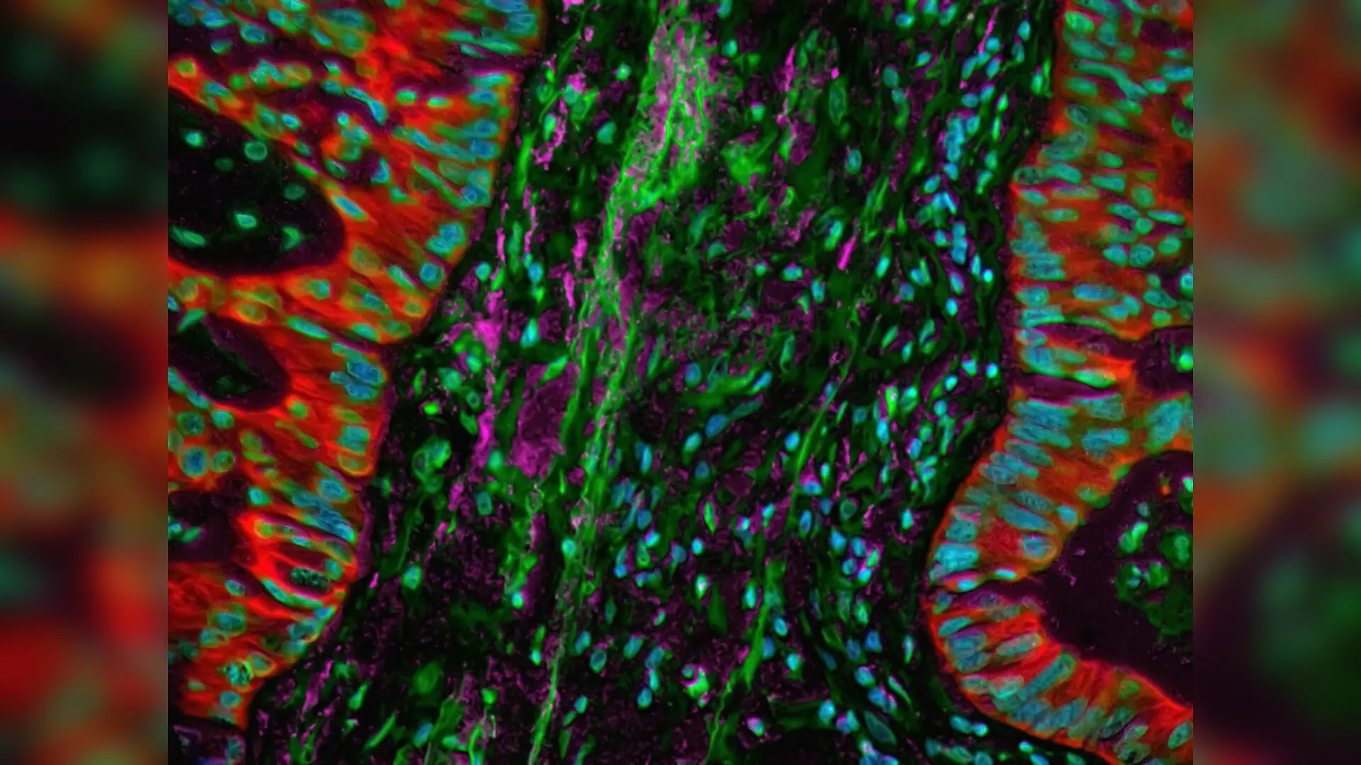
Scientists have mapped the first cross-species cellular atlas of the knee meniscus, revealing a hidden population of smooth muscle cells that sharply dwindle during joint disease. The comprehensive genetic investigation compares the tough, shock-absorbing cartilage tissue across mice, rats, beagles, goats, and humans to better understand why knees degenerate over time.
Decoding the Microscopic Blueprint Across Species
The meniscus is a crescent-shaped piece of fibrocartilage sitting between the thigh bone and shin bone, acting as the body’s primary shock absorber. When this vital tissue tears or wears down, it often triggers progressive joint failure and osteoarthritis. To understand its microscopic makeup, researchers previously relied on isolated human samples or single-animal models. By applying single-cell RNA sequencing, which analyzes the genetic activity of individual cells, the research team created an integrated cellular map of the meniscus across multiple mammalian species.
The analysis, published in Bone & Joint Research, identified nine distinct cell types shared across all examined mammals. These core populations include chondrocytes (cartilage-producing cells), fibroblasts ( connective tissue cells), and specialized fibrochondrocytes. However, their proportions varied widely depending on the species. Interestingly, the cellular landscape of rat meniscus closely mirrored that of human tissue, suggesting that rodents may serve as a more accurate laboratory model for human knee conditions than previously recognized.
The Disappearing Act of Smooth Muscle Cells in Diseased Joints
A surprising discovery in the cellular atlas was the consistent presence of actin alpha 2-positive smooth muscle cells, often referred to as ACTA2-positive cells, residing mainly in the outer surface zone of the meniscus. While smooth muscle cells typically line blood vessels to help control blood flow, these specialized cells appeared throughout non-vascular areas of the tissue. Through histological staining of human samples, the authors discovered that these protective smooth muscle cells experience a sharp reduction in patients suffering from osteoarthritis, rheumatoid arthritis, and discoid lateral meniscus, a congenital deformity.
Further laboratory experiments tracked these cells over time and investigated how they communicate with surrounding tissue. By cross-referencing genetic data, the team pinpointed three conserved genes enriched in these smooth muscle populations, naming SPARCL1, S1PR3, and ARHGAP6 as key regulatory candidates. Functional co-culture assays revealed that activating the S1PR3 signaling pathway directly stimulates neighboring meniscal cells to produce collagen type 1 alpha 1, a foundational protein for tissue strength and repair.
What These Cellular Shifts Mean for Future Knee Therapies
The identification of a universal cellular atlas highlights both striking similarities and important differences in how mammalian joints maintain themselves. While small rodents such as mice and rats harbor bone marrow-like compartments inside their menisci that are entirely absent in dogs, goats, and humans, the core structural blueprint remains remarkably conserved. The observation that smooth muscle populations vanish during degenerative joint diseases suggests they play a crucial role in maintaining structural integrity and mechanosensing.
Despite these promising insights, the authors noted several limitations in their work. Human sample sizes were modest and did not cover a wide distribution of exact ages, and the current functional validations relied heavily on pharmacological agents rather than direct genetic knockouts. Future investigations will need to determine whether targeting these specific signaling pathways or finding ways to replenish lost smooth muscle cells can halt or reverse meniscal degeneration, potentially opening the door to regenerative treatments for millions of people living with chronic knee pain.
The research was published in Bone & joint research on November 17, 2026.
This article has been fact checked for accuracy, with information verified against reputable sources. Learn more about us and our editorial process.
Last reviewed on .
Article history
- Latest version
Reference(s)
- Zhang, Mengyuan., et al. “Atlas of meniscal cells from different species and characterizing meniscal endothelial and smooth muscle cells.” Bone & Joint Research, vol. 15, no. 11, November 17, 2026, pp. 1374-1385. British Editorial Society of Bone & Joint Surgery, doi: 10.1302/2046-3758.1511.bjr-2026-0026.r2. <https://doi.org/10.1302/2046-3758.1511.bjr-2026-0026.r2>.
Cite this page:
- Posted by Elizabeth Taylor