Human Blood Stem Cells Reveal Surprising Clonal Dynamics in Mice
Environmental Science

Human Blood Stem Cells Reveal Surprising Clonal Dynamics in Mice

Tracking purified human stem and progenitor cells uncovers how conditioning regimens and serial transplants alter blood regeneration.
By William Moore
Published:
Email this Article
Human Blood Stem Cells Reveal Surprising Clonal Dynamics in Mice
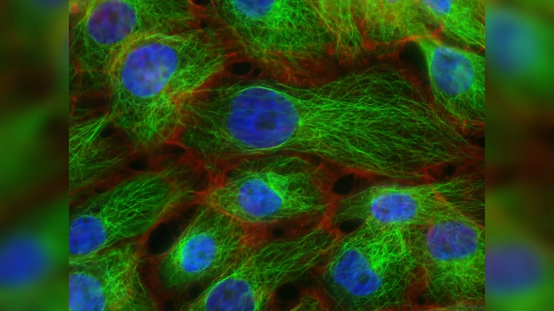
Photo by National Cancer Institute on Unsplash

When human blood stem cells are transplanted into animal models, they behave in surprisingly unexpected ways depending on the chemical or radiation treatments used to prepare the host. Researchers tracked thousands of individual human stem and progenitor cells inside specialized immunodeficient mice to observe how distinct cell populations rebuild the blood and immune system over time.

The study, published in NPJ Regenerative Medicine, was led by investigators at the University of Southern California including Mary Vergel Snodgrass, Jiya Eerdeng, Patrick Condie, Basia Gala, and senior author Rong Lu. The team investigated human hematopoietic stem cells (which give rise to lifelong blood and immune cells) and human multipotent progenitors (downstream cells with limited engraftment capacity) to untangle their precise roles in blood regeneration.

How Stem Cells Respond to Pre-Transplant Conditioning

Before bone marrow transplants, patients typically undergo pre-transplant conditioning regimens such as chemotherapy drugs like busulfan or total body irradiation to clear out existing cells and make room for donor tissue. To see how these treatments affect specific cell types, the researchers labeled 7,000 human stem cells and 18,000 progenitor cells with unique genetic barcodes before injecting them into NSGW41 mice that were either unconditioned, treated with busulfan, or exposed to irradiation.
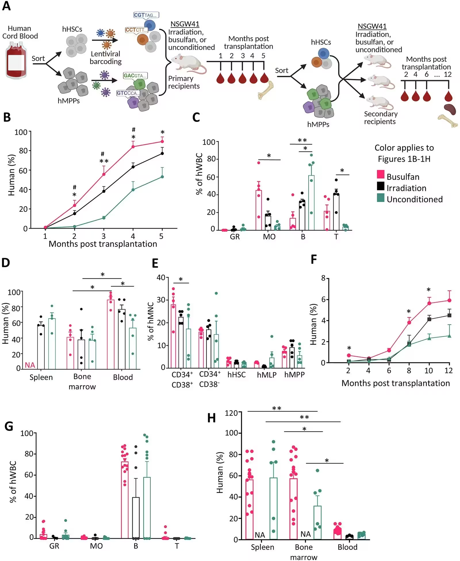
Fig. 1: Blood cell production by hHSPCs is influenced by pre-transplant conditioning. A Schematic overview of the experimental workflow. For primary transplantation, five mice were transplanted per conditioning group. For secondary transplantation, data were collected from 17 busulfan-conditioned, 11 unconditioned, and 6 irradiated recipients. B Percentage of human hematopoietic cells (hCD45+) in the peripheral blood of primary recipient mice over time. C Composition of human-derived white blood cells in the peripheral blood of primary recipients at 5 months post-transplantation. D Percentage of human hematopoietic cells in various tissues of the primary recipients at 5 months post-transplantation. E Frequency of human HSPCs among human mononuclear cells in the BM of the primary recipients at 5 months post-transplantation. F Percentage of human hematopoietic cells in the peripheral blood of secondary recipient mice. Significant differences were observed between the busulfan and unconditioned g
Blood cell production by hHSPCs is influenced by pre-transplant conditioning. A Schematic overview of the experimental workflow. For primary transplantation, five mice were transplanted per conditioning group. For secondary transplantation, data were collected from 17 busulfan-conditioned, 11 unconditioned, and 6 irradiated recipients. B Percentage of human hematopoietic cells (hCD45+) in the peripheral blood of primary recipient mice over time. C Composition of human-derived white blood cells in the peripheral blood of primary recipients at 5 months post-transplantation. D Percentage of human hematopoietic cells in various tissues of the primary recipients at 5 months post-transplantation. E Frequency of human HSPCs among human mononuclear cells in the BM of the primary recipients at 5 months post-transplantation. F Percentage of human hematopoietic cells in the peripheral blood of secondary recipient mice. Significant differences were observed between the busulfan and unconditioned g Credit: Mary Vergel Snodgrass et al. / NPJ Regenerative medicine

The results indicate that conditioning significantly boosted the overall abundance of human blood cells derived from the transplant, with busulfan producing the highest chimerism levels. However, the treatments also shifted lineage bias. Busulfan skewed cell differentiation toward monocytes, irradiation promoted T cell production, and the complete absence of conditioning heavily favored B cell lymphopoiesis. Interestingly, while peripheral blood counts changed dramatically, the total human chimerism residing directly inside the bone marrow and spleen remained largely unaffected by the choice of conditioning.

What Happens During Serial Transplantation

To test the long-term self-renewal capacity of these populations, the researchers harvested cells from primary mice five months after injection and transplanted them into secondary recipient mice. This secondary phase revealed a drastic drop in efficiency, with peripheral blood chimerism falling below 6 percent across all groups, compared to 50 to 80 percent in primary recipients.

Unexpectedly, clonal tracking showed that more individual clones contributed to blood production in secondary recipients than in primary ones. This points to the activation of previously quiescent or dormant stem cell clones responding to the stress of serial transplantation. Furthermore, secondary recipients frequently developed extramedullary hematopoiesis, a condition where blood cell production happens outside the bone marrow, such as in the spleen, lymph nodes, ovaries, and lungs.
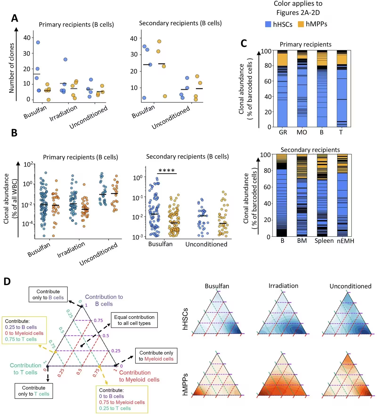
Fig. 2: Long-term multi-lineage differentiation of hHSCs and hMPPs. Comparing clonal contributions of hHSCs and hMPPs to the primary recipients 5 months post-transplantation and to the secondary recipients 12 months after secondary transplantation. A Number of hHSC and hMPP clones contributing to B cells in the peripheral blood of primary and secondary recipients. Each dot represents data from one recipient mouse. Black bars indicate the means. B Clonal contributions of hHSCs and hMPPs to B cells in the peripheral blood of primary and secondary recipients. Each dot represents a unique clone. Black bars indicate the medians; Mann–Whitney test. ****p < 0.0001. C Relative contributions of hHSC and hMPP clones to various hematopoietic cell types and tissues in the primary and secondary recipients. Each section within the columns represents one clone. Clones are ordered by their B cell contribution. D Lineage bias of hHSC and hMPP clones in primary recipients treated with busulfan, irradiation, or
Long-term multi-lineage differentiation of hHSCs and hMPPs. Comparing clonal contributions of hHSCs and hMPPs to the primary recipients 5 months post-transplantation and to the secondary recipients 12 months after secondary transplantation. A Number of hHSC and hMPP clones contributing to B cells in the peripheral blood of primary and secondary recipients. Each dot represents data from one recipient mouse. Black bars indicate the means. B Clonal contributions of hHSCs and hMPPs to B cells in the peripheral blood of primary and secondary recipients. Each dot represents a unique clone. Black bars indicate the medians; Mann–Whitney test. ****p < 0.0001. C Relative contributions of hHSC and hMPP clones to various hematopoietic cell types and tissues in the primary and secondary recipients. Each section within the columns represents one clone. Clones are ordered by their B cell contribution. D Lineage bias of hHSC and hMPP clones in primary recipients treated with busulfan, irradiation, or Credit: Mary Vergel Snodgrass et al. / NPJ Regenerative medicine

Limitations and Implications for Future Therapies

The authors noted several important limitations in their experimental design. Because preserving enough donor tissue for secondary transplants was critical, the team could not analyze tracking barcodes directly inside the bone marrow of primary recipients. Additionally, the mice used in the study provide limited support for human red blood cell production, and the human stem cells were sourced from umbilical cord blood rather than adult bone marrow.

Despite these caveats, the findings provide a high-resolution framework for understanding human blood regeneration at the single-clone level. By clarifying how conditioning drugs alter stem cell behavior and why extramedullary hematopoiesis occurs under stress, the work offers valuable insights that could eventually help optimize preclinical evaluations for stem cell and gene therapies.

The research was published in NPJ Regenerative medicine on November 17, 2026.

Fact Checked

This article has been fact checked for accuracy, with information verified against reputable sources. Learn more about us and our editorial process.

Last reviewed on .

Article history

  • Latest version

Reference(s)

  1. Vergel Snodgrass, Mary., et al. “Tracking human HSCs and MPPs in mice reveals distinct clonal dynamics and responses to pre-transplant conditioning.” npj Regenerative Medicine, vol. 11, no. 1, November 17, 2026 Springer Science and Business Media LLC, doi: 10.1038/s41536-026-00522-6. <https://doi.org/10.1038/s41536-026-00522-6>.

Cite this page:

Moore, William. “Human Blood Stem Cells Reveal Surprising Clonal Dynamics in Mice.” BioScience. BioScience ISSN 2521-5760, 20 November 2026. <https://www.bioscience.com.pk/en/subject/environmental-science/tracking-human-hscs-and-mpps-in-mice-reveals-distinct-clonal-dynamics-and-responses-to-pre-transplant-conditioning>. Moore, W. (2026, November 20). “Human Blood Stem Cells Reveal Surprising Clonal Dynamics in Mice.” BioScience. ISSN 2521-5760. Retrieved November 20, 2026 from https://www.bioscience.com.pk/en/subject/environmental-science/tracking-human-hscs-and-mpps-in-mice-reveals-distinct-clonal-dynamics-and-responses-to-pre-transplant-conditioning Moore, William. “Human Blood Stem Cells Reveal Surprising Clonal Dynamics in Mice.” BioScience. ISSN 2521-5760. https://www.bioscience.com.pk/en/subject/environmental-science/tracking-human-hscs-and-mpps-in-mice-reveals-distinct-clonal-dynamics-and-responses-to-pre-transplant-conditioning (accessed November 20, 2026).
End of the article