New Fluorescent Sensor Lights Up Cancer Cells by Targeting Hidden Sulfur Biomarkers
A newly synthesized water-soluble probe exploits a unique molecular transformation to distinguish malignant tissues from healthy cells under a microscope.
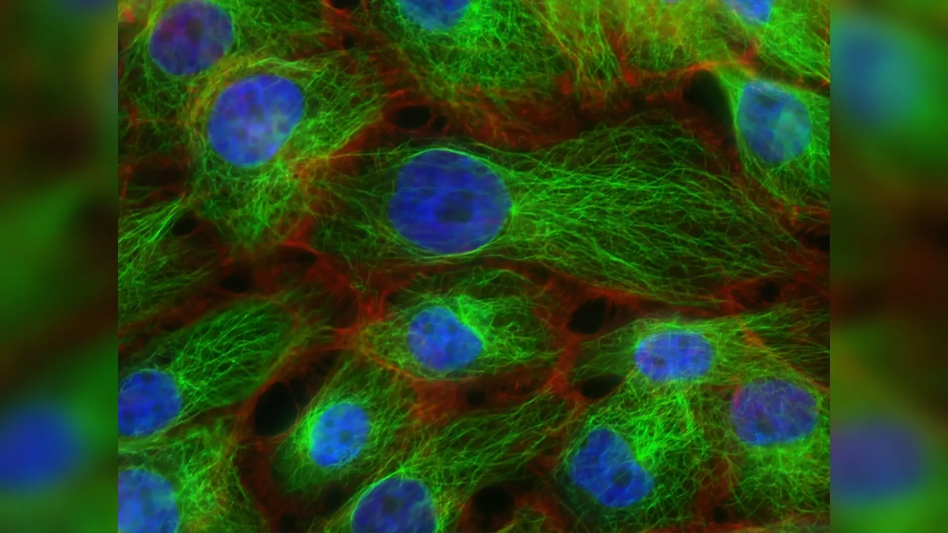
A newly designed fluorescent molecule lights up green when it encounters the reactive sulfur-containing groups found abundantly in tumor cells, offering a fresh way to spot the difference between malignant and healthy tissues under a microscope. The water-soluble probe, named P3, was successfully tested on human lung and colon cancer cell lines, where it produced a stark contrast in fluorescence compared to their healthy epithelial counterparts.
Sulfur-bearing molecules known as thiols, including the cellular antioxidant glutathione and the amino acid cysteine, are essential for keeping cells in chemical balance. However, cancer cells frequently accumulate higher levels of these compounds to protect themselves against oxidative stress and neutralize the effects of chemotherapy drugs. Detecting these biomarkers accurately has traditionally required expensive and labor-intensive laboratory equipment such as mass spectrometers or high-performance liquid chromatography systems.
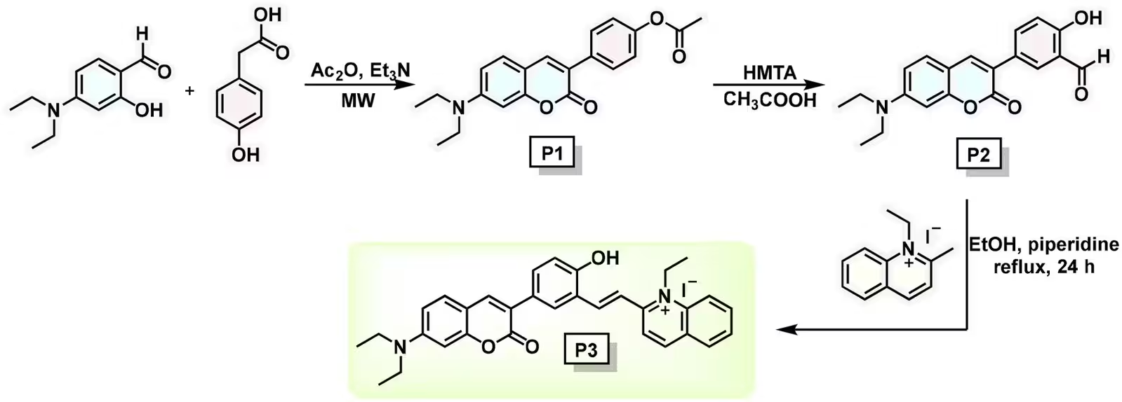
To overcome those hurdles, a team of researchers synthesized a hybrid chemical structure combining a coumarin derivative with a quinolinium salt. Details of the creation and testing procedure were published in The Analyst. The resulting compound exhibits a large Stokes shift of approximately 90 nanometers, meaning its emitted green light is well separated from the light used to excite it, which reduces background noise and sharpens microscopic images.
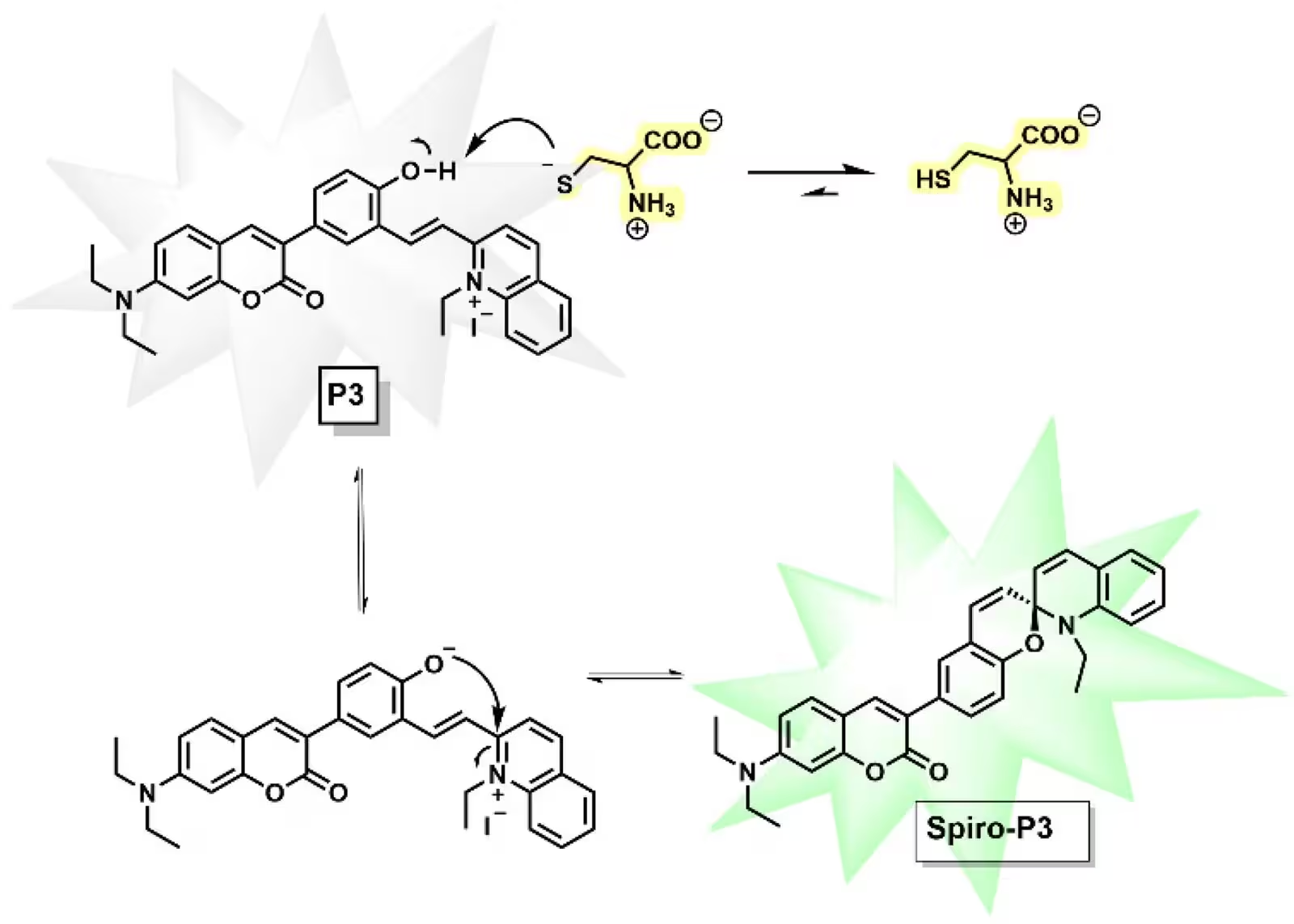
How the Fluorescent Switch Operates Inside Cells
The sensor relies on a chemical transformation that turns an extended, conjugated molecular structure into a spirocyclic form. When a free thiol comes into contact with the probe, it acts as a chemical base and abstracts a proton from the molecule’s phenolic hydroxyl group. This triggers an intramolecular attack that changes the electron distribution, locking the molecule into a rigid ring framework that prevents non-radiative energy loss and turns on the green fluorescence.
Laboratory tests showed that the probe responds selectively to thiols within ten minutes in a near-neutral buffer solution mimicking physiological pH. Non-thiol amino acids, vitamins, and metal ions generated less than a ten percent relative signal change, confirming the high specificity of the sensor. The detection limits for cysteine and glutathione reached low micromolar concentrations of 5.32 and 5.94 micromolars, respectively.
Potential Uses Beyond Cancer Diagnostics
Beyond identifying cancer cells, the research team explored whether the sensor could monitor structural changes in proteins and analyze commercial health products. When introduced to bovine serum albumin, a model protein containing a single accessible thiol group, the probe produced a bright fluorescence enhancement and a color shift from green to blue, likely because the molecule became trapped inside the protein’s hydrophobic pockets. The sensor also successfully evaluated over-the-counter dietary supplements containing cysteine, matching the accuracy of traditional testing methods.
In live-cell imaging experiments, human colorectal adenocarcinoma cells and lung carcinoma cells displayed intense green fluorescence after a brief ten-minute incubation with the probe. Meanwhile, healthy human colon and lung epithelial cells showed virtually no signal under identical conditions. Confocal microscopy further revealed that the probe accumulates in a granular, punctate pattern within the cytoplasm.
The authors note that the reaction is part of a reversible equilibrium rather than a permanent chemical destruction, opening doors for future real-time tracking of redox fluctuations in complex biological systems. While further biological validation is necessary before clinical implementation, the sensor provides a versatile, cost-effective platform for investigating redox-related diseases and visualizing intracellular biomarkers.
The research was published in The Analyst on November 13, 2026.
This article has been fact checked for accuracy, with information verified against reputable sources. Learn more about us and our editorial process.
Last reviewed on .
Article history
- Latest version
Reference(s)
- Abdulsalam Abdulsalam, Mohammed Adnan., et al. “Coumarin-quinolinium based fluorescent probe for free thiol detection and cancer cell imaging.” The Analyst, January 1, 2026 Royal Society of Chemistry (RSC), doi: 10.1039/d6an00818f. <https://doi.org/10.1039/d6an00818f>.
Cite this page:
- Posted by William Moore