Popular Vaccine Stabilizer Triggers Unwanted Immune Response
Biotechnology

Popular Vaccine Stabilizer Triggers Unwanted Immune Response

Adding sugar shields to the viral foldon domain reduces binding for some antibodies, but fails to stop unwanted immune activation in naive subjects.
By Rohan Kumar
Published:
Email this Article
Popular Vaccine Stabilizer Triggers Unwanted Immune Response
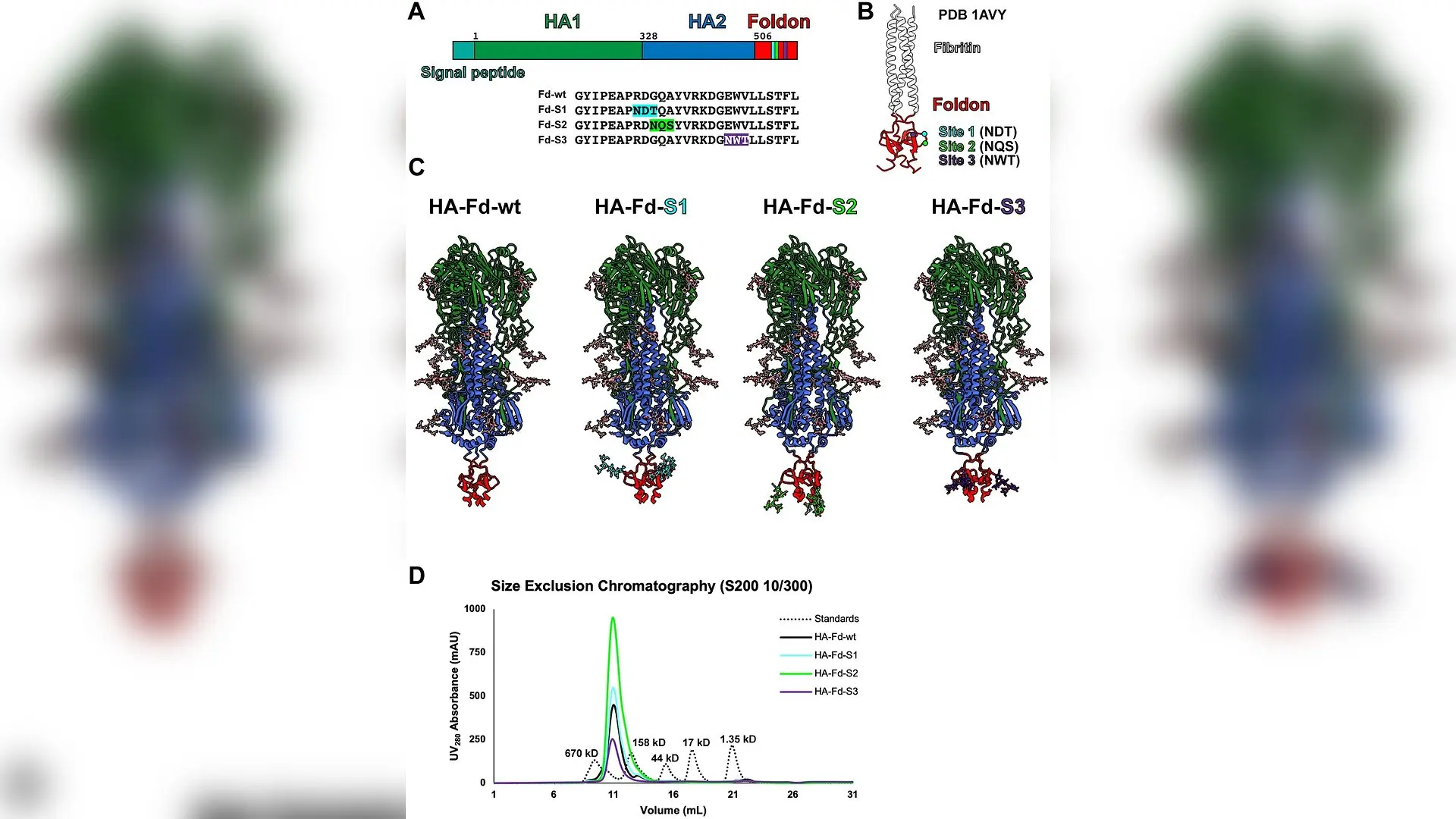
Design, expression, and purification of hemagglutinin Foldon mutants. (A) Construct design of the HA-Foldon mutants. The relative positions of sites 1, 2 and 3 in the protein sequence are marked as cyan, bright green, and purple bars, respectively. The sequences of the different Foldon versions are shown. The C-terminal AviTag and hexahistidine tag are removed for simplicity. (B) Crystal structure (PDB 1AVY) of the fibritin protein used for structure-based protein design. The approximate locations of the mutation sites within the structure are denoted by dots, colored as in (A). (C) Predicted models of the wildtype and mutant protein constructs from AlphaFold3 containing mammalian glycosylation trees, with complex branching excluded for simplicity. Protein regions are colored as in (A). Glycan residues linked to the HA are shown in gray, while those on the Foldon are color-coded based on the mutation site. The residues comprising the AviTags and hexahistidine tags have been hidden for John V Dzimianski et al. / PloS one

Protein subunit and mRNA vaccines rely on specialized structural domains to hold fragile viral proteins in the correct shape, but a new study reveals that one of the most popular stabilizing tools triggers unexpected immune blind spots. Researchers investigated the Foldon trimerization domain, a tiny protein segment derived from the T4 bacteriophage that holds together viral antigens in FDA-approved vaccines such as AREXVY and ABRYSVO for respiratory syncytial virus.

While the Foldon domain successfully stabilizes vital vaccine antigens, previous clinical trials showed it also provokes unwanted off-target antibody responses in humans and animals. To solve this problem, a team led by scientists at the University of California Santa Cruz and the University of Georgia set out to build an invisible molecular shield around the domain by adding sugar molecules through genetic engineering. The findings were published in PLOS One, detailing both the potential and the strict structural limits of modifying viral subunit components.

How Scientists Tried to Hide the Foldon Domain Using Engineered Sugars

The research team focused on modifying a computationally optimized broadly reactive antigen, known as a COBRA influenza hemagglutinin protein, by appending the Foldon domain to its tail end. Using structural models of the T4 fibritin protein, the authors selected three solvent-exposed sites, designated as Site 1, Site 2, and Site 3, to introduce specific amino acid sequences that signal cells to attach N-linked glycan carbohydrates. These sugary additions are naturally used by host cells to coat proteins and evade detection by the immune system.
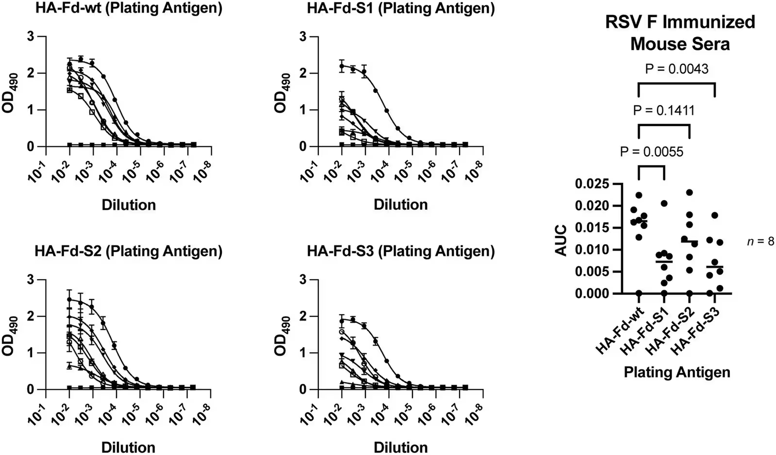
Fig 2: ELISA data with HA-Foldon antigens and RSV immunized mouse sera. Plates were coated with the indicated antigen and tested across the same sera samples. Technical duplicates of eight biological replicates were analyzed in Prism by fitting the data to a nonlinear 4 parameter model, followed by calculation of the area under the curve (AUC). Each data point represents the mean of the technical duplicates, with error bars indicating the standard deviation. A repeated measures one-way ANOVA with Dunnett’s multiple comparisons test was performed to track the changes in each biological replicate from the plate coated with wildtype antigen compared to each plate coated with one of the engineered antigens. One biological replicate showed no response in all of the assays.
ELISA data with HA-Foldon antigens and RSV immunized mouse sera. Plates were coated with the indicated antigen and tested across the same sera samples. Technical duplicates of eight biological replicates were analyzed in Prism by fitting the data to a nonlinear 4 parameter model, followed by calculation of the area under the curve (AUC). Each data point represents the mean of the technical duplicates, with error bars indicating the standard deviation. A repeated measures one-way ANOVA with Dunnett’s multiple comparisons test was performed to track the changes in each biological replicate from the plate coated with wildtype antigen compared to each plate coated with one of the engineered antigens. One biological replicate showed no response in all of the assays. Credit: John V Dzimianski et al. / PloS one

When the researchers expressed these redesigned proteins in mammalian Chinese hamster ovary cells, single-site mutations successfully formed stable trimers that closely mirrored the unmodified wild-type protein. However, attempting to combine two of these glycosylation sites into a single protein caused the trimeric structure to fall apart into monomers. This outcome pointed to a rigid boundary in how much genetic tampering the Foldon domain can tolerate before its core stability collapses.

Testing the Shield Against Monoclonal Antibodies and Mouse Sera

The investigators then tested whether the added sugar coatings could successfully block antibody recognition using enzyme-linked immunosorbent assays and biolayer interferometry. When exposed to sera from mice previously immunized with respiratory syncytial virus antigens containing a wild-type Foldon, the single-site glycan mutants at Site 1 and Site 3 demonstrated a statistically significant reduction in binding reactivity.
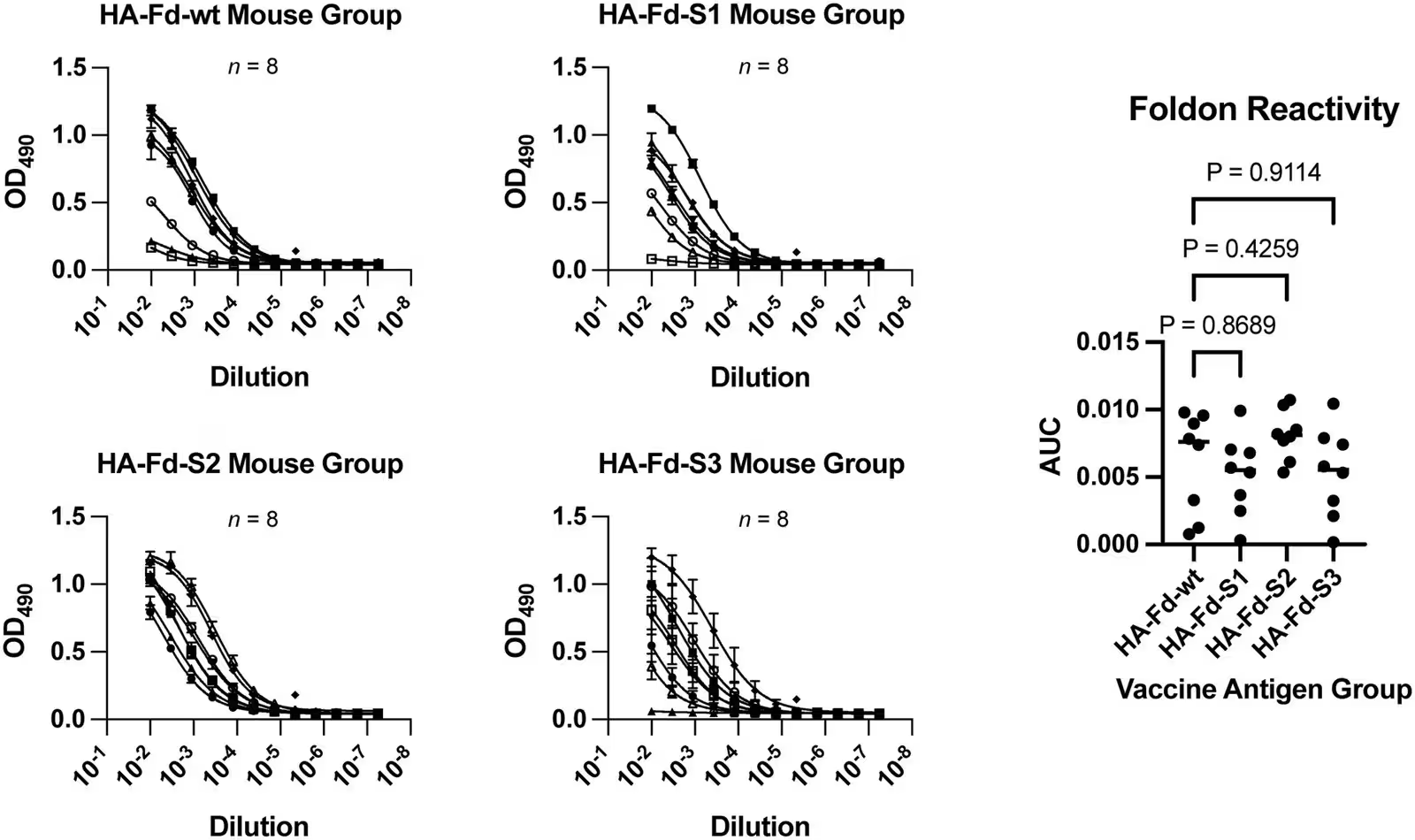
Fig 3: ELISA data with recombinant Foldon and HA-Foldon immunized mouse groups. The plates were coated with Foldon and tested with sera obtained from mouse groups immunized with four different HA-Foldon antigens. The assay with each group was run with technical duplicates with eight biological replicates. Each data point represents the mean of technical duplicates with error bars for the standard deviation. The data were analyzed in Prism by fitting the data to a nonlinear 4 parameter model, followed by calculation of the area under the curve (AUC). An ordinary one-way ANOVA with Dunnett’s multiple comparisons test was performed to assess the differences of each Foldon mutant vaccine group relative to the control group vaccinated with wildtype antigen.
ELISA data with recombinant Foldon and HA-Foldon immunized mouse groups. The plates were coated with Foldon and tested with sera obtained from mouse groups immunized with four different HA-Foldon antigens. The assay with each group was run with technical duplicates with eight biological replicates. Each data point represents the mean of technical duplicates with error bars for the standard deviation. The data were analyzed in Prism by fitting the data to a nonlinear 4 parameter model, followed by calculation of the area under the curve (AUC). An ordinary one-way ANOVA with Dunnett’s multiple comparisons test was performed to assess the differences of each Foldon mutant vaccine group relative to the control group vaccinated with wildtype antigen. Credit: John V Dzimianski et al. / PloS one

However, when the team vaccinated a new group of naive mice with the glycan-shielded antigens, the animals still generated robust antibody responses directly targeting the Foldon domain. The sugar-modified vaccines failed to significantly lower overall Foldon immunogenicity in subjects with no prior exposure. Further biochemical analyses utilizing mass spectrometry and enzymatic deglycosylation revealed that the sugar occupancy at the target sites was less than 50 percent. Moreover, mutating the underlying amino acids alone accounted for much of the altered antibody binding, independent of the attached sugars.

What These Limits Mean for Future Vaccine Design

As biotechnology researchers continue to develop advanced subunit vaccines for viral pathogens, managing off-target immunity remains a critical priority. The study highlights that while introducing glycosylation motifs can alter local protein antigenicity, single-site shields are currently insufficient to prevent the immune system from recognizing the underlying Foldon scaffold. The authors conclude that alternative strategies, such as developing entirely self-trimerizing viral antigens without exogenous helper domains, will likely be necessary to prevent unintended immune reactions in future clinical applications.

The research was published in PloS one on January 1, 2026.

Fact Checked

This article has been fact checked for accuracy, with information verified against reputable sources. Learn more about us and our editorial process.

Last reviewed on .

Article history

  • Latest version

Reference(s)

  1. Dzimianski, John V.., et al. “Immunogenicity of the Foldon trimerization domain and efforts to shield it.” PLOS One, vol. 21, no. 11, November 13, 2026, pp. e0362571 Public Library of Science (PLoS), doi: 10.1371/journal.pone.0362571. <https://doi.org/10.1371/journal.pone.0362571>.

Cite this page:

Kumar, Rohan. “Popular Vaccine Stabilizer Triggers Unwanted Immune Response.” BioScience. BioScience ISSN 2521-5760, 16 November 2026. <https://www.bioscience.com.pk/en/subject/biotechnology/immunogenicity-of-the-foldon-trimerization-domain-and-efforts-to-shield-it>. Kumar, R. (2026, November 16). “Popular Vaccine Stabilizer Triggers Unwanted Immune Response.” BioScience. ISSN 2521-5760. Retrieved November 16, 2026 from https://www.bioscience.com.pk/en/subject/biotechnology/immunogenicity-of-the-foldon-trimerization-domain-and-efforts-to-shield-it Kumar, Rohan. “Popular Vaccine Stabilizer Triggers Unwanted Immune Response.” BioScience. ISSN 2521-5760. https://www.bioscience.com.pk/en/subject/biotechnology/immunogenicity-of-the-foldon-trimerization-domain-and-efforts-to-shield-it (accessed November 16, 2026).
End of the article