Hidden Body Map Discovered Inside the Brains Secondary Motor Cortex
Biotechnology

Hidden Body Map Discovered Inside the Brains Secondary Motor Cortex

Resting brain activity reveals a precise topographical layout that helps mammals coordinate complex movements.
By Robert Brown
Published:
Email this Article
Hidden Body Map Discovered Inside the Brains Secondary Motor Cortex
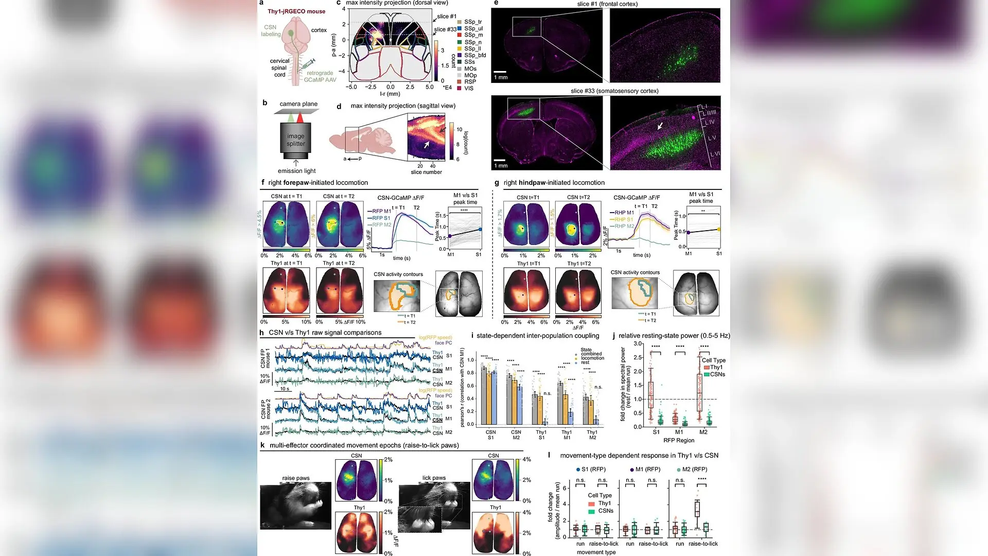
Thy1 and CSN populations exhibit distinct functional roles across diverse behaviors. a Retrograde AAV-GCaMP injection into cervical (forepaw) or lumbar (hindpaw) spinal cord. b Image splitter for dual-fluorescence imaging in Thy1-jRGECO mice. c Cortex-restricted max intensity projection (MIP) of GFP-labeled forelimb CSNs from histology sections, overlaid on the Allen Brain Atlas. d Sagittal MIP; arrows, spinally projecting CSNs (a, anterior; p, posterior). e Coronal sections from (c): slice #1 (frontal cortex, top) and #33 (somatosensory cortex, bottom); CSNs, green; DAPI, magenta; inset zoom, layers L I–L VI; white arrow, thinning of layer IV. Illustrative example: same features were observed in a second mouse. f Simultaneous imaging of forepaw CSNs (GCaMP) and Thy1 (jRGECO) during right forepaw-initiated locomotion, shown for one representative mouse of two (n = 113 epochs): Cortical maps show averaged CSN (top) and Thy1 (bottom) ∆F/F maps at T1 (left) and T2 (right); Plot shows aver Weihao Xu et al. / Nature communications

Brain mapping researchers have uncovered a hidden, highly organized map within the mouse secondary motor cortex that mirrors the body layout found in sensory areas. The discovery resolves a long-standing challenge in neuroscience by revealing how the brain coordinates complex movements without getting tangled in sensory noise.

Mapping the motor cortex has historically proven difficult because physical movements are invariably accompanied by sensory feedback and postural adjustments. To bypass this barrier, researchers led by Elizabeth Hillman and their team at Columbia University capitalized on moments of quiet rest rather than active movement. Using advanced optical imaging techniques published in Nature Communications, the team tracked cellular activity across the dorsal cortex of awake mice over multiple recording sessions.

How Resting Brain Fluctuations Reveal Hidden Maps

During periods of quiet rest, the mammalian brain exhibits spontaneous, heterogeneous neuronal fluctuations that were previously thought to be random background noise. The authors found that these resting oscillations are actually selectively synchronized between specific sub-regions of the primary sensory cortex and the secondary motor cortex. When the mice began moving, these distinct resting fluctuations abruptly quenched. By analyzing the correlation patterns during rest, the team could isolate functional connections without the confounding signals generated by active muscle contractions.
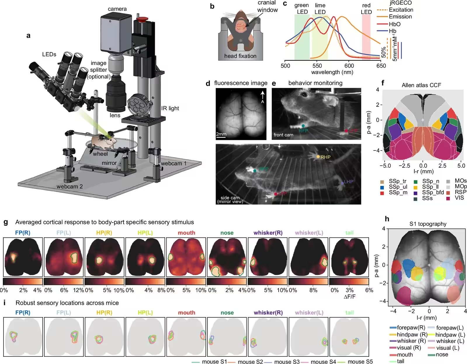
Fig. 1: Mapping sensory (S1) topography in the mouse dorsal cortex using wide-field optical mapping. a WFOM system diagram showing strobing LEDs illuminating the cortical surface in various wavelengths. Mice were head-fixed under the camera on a horizontal transparent wheel. Behaviors of the mice were monitored under infrared illumination using two webcams. An optional image splitter was used in two-color fluorescence imaging. CAD models of Thorlabs optomechanical components used with permission. b Illustration of the implanted headplate over a thinned skull cranial window. c The jRGECO1a fluorescence excitation, emission spectra, overlaid with the absorption spectra of oxy-hemoglobin (HbO), deoxy-hemoglobin (HbR), as well as the bands indicating the wavelengths of the illuminating LEDs used in WFOM (green, lime and red). d The dorsal view of the jRGECO1a fluorescence image through the mouse thinned skull. A: anterior. e Behavior monitoring from the front webcam (Top) and the side webcam captu
Mapping sensory (S1) topography in the mouse dorsal cortex using wide-field optical mapping. a WFOM system diagram showing strobing LEDs illuminating the cortical surface in various wavelengths. Mice were head-fixed under the camera on a horizontal transparent wheel. Behaviors of the mice were monitored under infrared illumination using two webcams. An optional image splitter was used in two-color fluorescence imaging. CAD models of Thorlabs optomechanical components used with permission. b Illustration of the implanted headplate over a thinned skull cranial window. c The jRGECO1a fluorescence excitation, emission spectra, overlaid with the absorption spectra of oxy-hemoglobin (HbO), deoxy-hemoglobin (HbR), as well as the bands indicating the wavelengths of the illuminating LEDs used in WFOM (green, lime and red). d The dorsal view of the jRGECO1a fluorescence image through the mouse thinned skull. A: anterior. e Behavior monitoring from the front webcam (Top) and the side webcam captu Credit: Weihao Xu et al. / Nature communications

The resulting functional maps demonstrated that the secondary motor cortex contains a precise, body-part-specific topography. Distinct clusters corresponded to the forepaws, hindpaws, whiskers, and mouth, arranging themselves in a layout that structurally mirrored the organization of the primary somatosensory cortex. Structural tracing data using the Allen Mouse Brain Connectivity Atlas confirmed that physical axonal projections travel from sensory areas through deep cortical layers to target these exact homologous sub-regions in the motor cortex.

Distinguishing Motor Output From Cortical Preparation

To confirm that these resting fluctuations did not represent actual motor output, the investigators used retrograde viral tracers to label corticospinal neurons, which are the primary cells that send direct commands from the brain to the spinal cord. Simultaneous dual-color imaging revealed that while pan-neuronal populations showed strong synchronized fluctuations during rest, the corticospinal motor output neurons remained completely silent and near baseline during these resting periods.
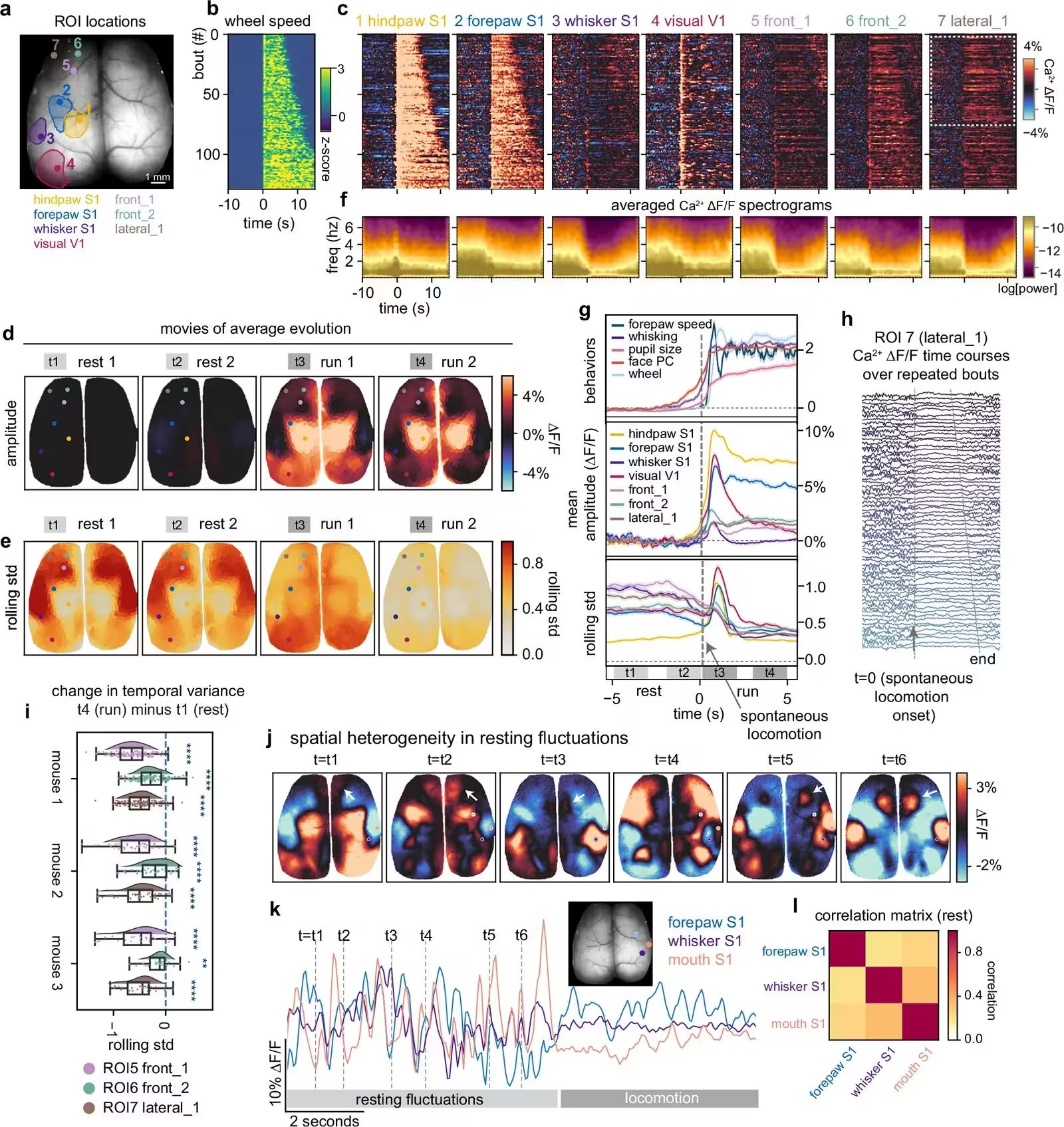
Fig. 2: Cortical representations of spontaneous behaviors. a The selected ROIs, including V1/S1 and additional frontal cortical regions (front_1, front_2, and lateral_1), are overlaid on the Thy1-jRGECO1a fluorescence image. Sensory mapping contours are shown for the S1/V1 regions. Panels (b–h) show data from a representative mouse (n = 130 rest-to-locomotion bouts); panel (i) aggregates three mice. b Locomotion behavior in 130 extracted rest-to-locomotion bouts. The heatmap displays the wheel speed for each epoch. The onset of each locomotion bout is aligned at time t = 0, with a minimum of 10 seconds of rest preceding the locomotion onset. The durations of locomotion span from 5 to 15 seconds in all extracted bouts. c, Corresponding neural activation (ΔΔF/F) for 7 selected ROIs during 130 extracted bouts. d, e Maps of the average neural activation ΔΔF/F and moving window standard deviation for 4 time windows (rest 1, rest 2, run 1, run 2) indicated in the lower panel of (g) (n = 130 bouts).
Cortical representations of spontaneous behaviors. a The selected ROIs, including V1/S1 and additional frontal cortical regions (front_1, front_2, and lateral_1), are overlaid on the Thy1-jRGECO1a fluorescence image. Sensory mapping contours are shown for the S1/V1 regions. Panels (b–h) show data from a representative mouse (n = 130 rest-to-locomotion bouts); panel (i) aggregates three mice. b Locomotion behavior in 130 extracted rest-to-locomotion bouts. The heatmap displays the wheel speed for each epoch. The onset of each locomotion bout is aligned at time t = 0, with a minimum of 10 seconds of rest preceding the locomotion onset. The durations of locomotion span from 5 to 15 seconds in all extracted bouts. c, Corresponding neural activation (ΔΔF/F) for 7 selected ROIs during 130 extracted bouts. d, e Maps of the average neural activation ΔΔF/F and moving window standard deviation for 4 time windows (rest 1, rest 2, run 1, run 2) indicated in the lower panel of (g) (n = 130 bouts). Credit: Weihao Xu et al. / Nature communications

Further analysis of different behavioral types showed that the secondary motor cortex engages most strongly during complex, multi-effector coordinated movements, such as a sequence involving both wheel locomotion and raising or licking paws. During simple, rhythmic locomotion, the primary motor and sensory regions dominated activation, whereas the secondary motor areas showed lower-amplitude participation. This task-dependent engagement points to a role in overarching motor coordination, sensory integration, and gating unwanted movements rather than directly driving basic muscle contractions.

What This Means for Future Motor System Research

The findings provide a clear framework for interpreting intrinsic sensorimotor dynamics and help explain why past motor cortex stimulation studies yielded conflicting results. Because traditional experiments often average movement-locked activity across trials, they frequently filter out the intrinsic cortical dynamics that occur during resting states. The authors note that while these methods successfully mapped the mouse cortex using wide-field optical mapping, future investigations will need to combine high-speed in-vivo microscopy and single-cell resolution to fully decode how mammalian brains process latent cognitive signals and plan intricate physical actions.

The research was published in Nature communications on November 13, 2026.

Fact Checked

This article has been fact checked for accuracy, with information verified against reputable sources. Learn more about us and our editorial process.

Last reviewed on .

Article history

  • Latest version

Reference(s)

  1. Xu, Weihao., et al. “Intrinsic neural dynamics reveal a body-part-specific topography in mouse secondary motor cortex.” Nature Communications, vol. 17, no. 1, November 13, 2026 Springer Science and Business Media LLC, doi: 10.1038/s41467-026-78900-3. <https://doi.org/10.1038/s41467-026-78900-3>.

Cite this page:

Brown, Robert. “Hidden Body Map Discovered Inside the Brains Secondary Motor Cortex.” BioScience. BioScience ISSN 2521-5760, 16 November 2026. <https://www.bioscience.com.pk/en/subject/biotechnology/intrinsic-neural-dynamics-reveal-a-body-part-specific-topography-in-mouse-secondary-motor-cortex>. Brown, R. (2026, November 16). “Hidden Body Map Discovered Inside the Brains Secondary Motor Cortex.” BioScience. ISSN 2521-5760. Retrieved November 16, 2026 from https://www.bioscience.com.pk/en/subject/biotechnology/intrinsic-neural-dynamics-reveal-a-body-part-specific-topography-in-mouse-secondary-motor-cortex Brown, Robert. “Hidden Body Map Discovered Inside the Brains Secondary Motor Cortex.” BioScience. ISSN 2521-5760. https://www.bioscience.com.pk/en/subject/biotechnology/intrinsic-neural-dynamics-reveal-a-body-part-specific-topography-in-mouse-secondary-motor-cortex (accessed November 16, 2026).
End of the article