Math Tool Reveals Why One-Third of Biosensor Receptors Fail to Detect Targets
A novel distribution model resolves hidden electron-transfer states in aptamer sensors, shedding light on molecular binding mechanisms.
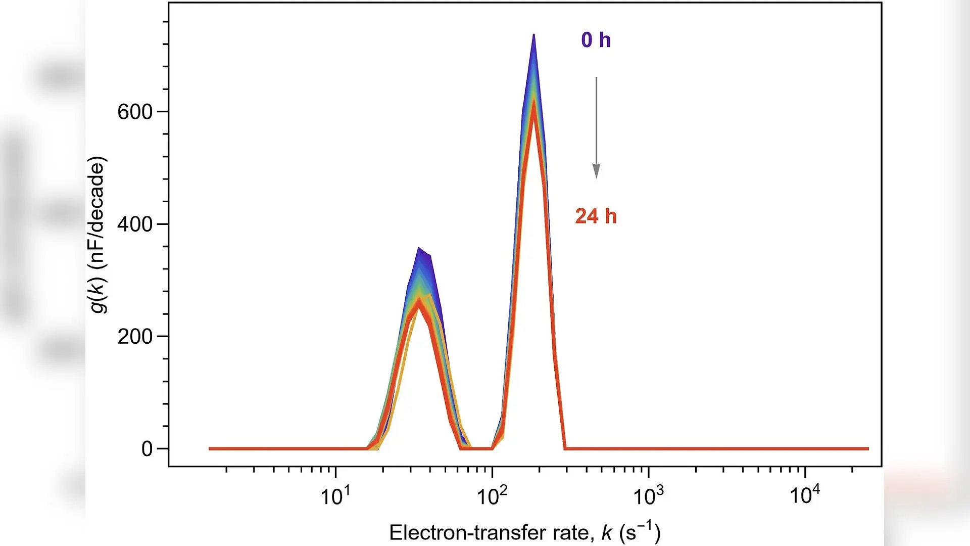
A new mathematical technique resolves hidden structural changes in electrochemical sensors, revealing why roughly one-third of surface-bound molecular receptors fail to respond to targets.
Electrochemical aptamer-based biosensors rely on molecular recognition elements tethered to an electrode surface to detect specific analytes in real time. These devices have advanced toward clinical diagnostic applications, yet understanding the precise physical mechanisms governing their signal generation has remained challenging. Traditional analytical methods often blur distinct kinetic pathways into a single averaged measurement, obscuring the true molecular behavior occurring at the solid-liquid interface.
To address this limitation, researchers applied an advanced mathematical approach called the Distribution of Capacitive Times model to parse complex electrical responses. The study, published in the Journal of the Electrochemical Society by Bob M. Lansdorp and colleagues, demonstrates that this method can accurately separate overlapping electron-transfer processes in surface-immobilized redox systems without relying on conventional approximations that violate physical boundaries.
How Maxwell Ladders Reveal Hidden Molecular States
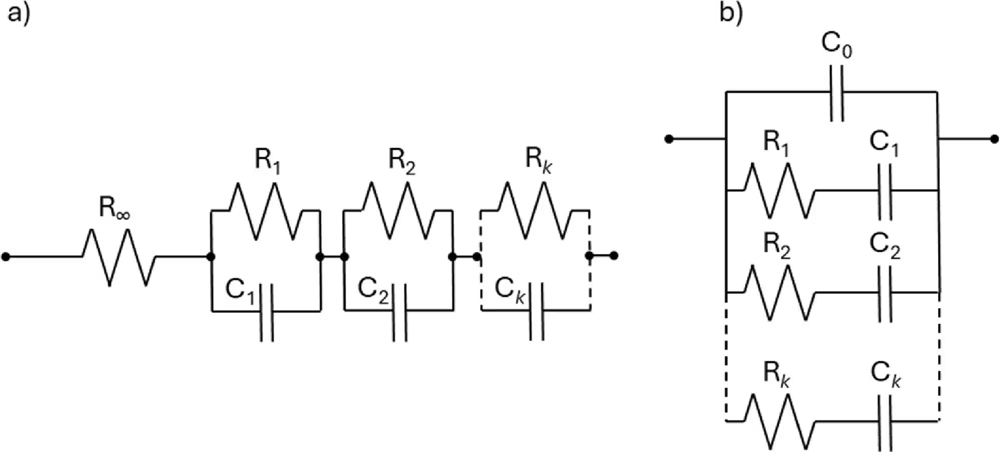
Standard electrochemical impedance spectroscopy measures how a system responds to small alternating voltage signals across a wide frequency range. Investigators previously interpreted these spectra using methods designed for batteries and fuel cells, which assume that electrical current flows sequentially through stacked material layers. However, sensors functionalized with tethered redox reporters feature a blocking molecular layer that creates an infinite impedance at low frequencies, rendering traditional sequential models invalid.
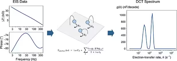
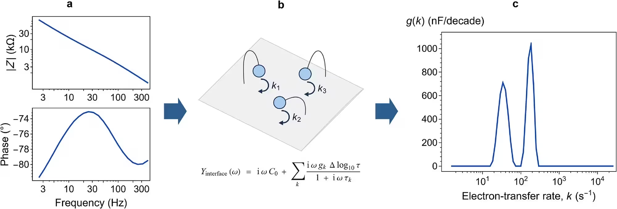
The research team utilized a parallel-connected circuit framework known as a Maxwell ladder, which matches the physical reality of surface-immobilized molecules sharing a single insulating interface. By implementing a linear-algebra-based numerical technique combined with Tikhonov regularization, the authors converted raw impedance spectra into high-resolution capacitive distribution profiles. This approach mathematically isolates distinct populations of molecules characterized by different electron-transfer rates, yielding global minimum solutions without depending on arbitrary initial estimates.
Unmasking Two Distinct Populations in Vancomycin Biosensors
When testing a biosensor designed to detect the antibiotic vancomycin, the analysis uncovered two distinct electron-transfer rate populations rather than a single continuous distribution. One population operated at a slow characteristic rate of approximately thirty-five inverse seconds, while the other operated at a fast rate of roughly one hundred eighty inverse seconds. These specific rates remained remarkably constant as target concentrations varied from zero to one millimolar.
Instead of the individual reaction rates shifting, titration experiments showed that the relative occupancy of each state evolved dynamically as more vancomycin bound to the sensor. As target concentrations increased, a progressively larger fraction of the molecular receptors shifted into the rapid electron-transfer state. This discovery strongly supports a conformational switching mechanism where molecules naturally fluctuate between states even before target capture occurs, rather than a model where binding directly induces the energetic changes.
Quantifying Folding Incompetence on Sensor Surfaces
When the team attempted to fit the concentration-dependent data using a standard three-state biophysical model, the experimental fractions plateaued near sixty percent rather than approaching full saturation. To account for this discrepancy, the authors introduced a fourth category representing non-folding aptamers that remain permanently incapable of structural reconfiguration due to steric crowding, close packing with neighboring molecules, or entrapment within microscopic crevices on the rough gold electrode.
Incorporating this non-folding population into the analytical framework yielded an exceptional agreement with experimental observations across multiple independent electrodes. The model produced a quantitative estimate indicating that approximately thirty-two percent of the aptamers populated on the sensor surface are structurally hindered from responding to their target. Recognizing and quantifying this inactive fraction provides a clear physical target for future engineering efforts aimed at improving sensor sensitivity and signal gain.
Broader Implications for Surface Electrochemistry
The successful deployment of capacitive timescale distributions highlights the critical importance of matching mathematical kernels to the actual physical properties of electrochemical interfaces. Accounting for series solution resistance prior to executing the inversion algorithm proved essential for resolving the dual peaks accurately. Without isolating the solution resistance, the distinct kinetic populations merged into unresolved broad features.
While the current work focused specifically on a vancomycin biosensor, the underlying analytical methodology holds broad potential for investigating other surface-immobilized redox reactions. Researchers anticipate that adapting these distributive analysis tools will enhance the mechanistic characterization of diverse electrochemical systems, ranging from advanced molecular sensors to catalytic interfaces and corrosion protection layers.
The research was published in Journal of the Electrochemical Society on November 13, 2026.
This article has been fact checked for accuracy, with information verified against reputable sources. Learn more about us and our editorial process.
Last reviewed on .
Article history
- Latest version
Reference(s)
- Lansdorp, Bob M.., et al. “Resolving Electron-Transfer Heterogeneity in Electrochemical Aptamer-Based Sensors by Distribution of Capacitive Times.” Journal of The Electrochemical Society, vol. 173, no. 21, November 12, 2026, pp. 216505 The Electrochemical Society, doi: 10.1149/1945-7111/aeb765. <https://doi.org/10.1149/1945-7111/aeb765>.
Cite this page:
- Posted by Robert Brown