Silk Protein Coating Transforms 3D-Printed Bone Scaffolds
Chemical modification alters plastic surfaces to boost wettability and cell adhesion without destroying delicate pore architectures.“, _comment: ”meta_description, meta_keywords, tags, publication_name
Engineers are trying to overcome the stubborn slickness of medical plastics that usually repel living cells, and a new laboratory study reveals how coating 3D-printed scaffolds with natural silk proteins changes everything. Bone tissue engineering relies heavily on artificial structures that give new bone cells a place to grow, but standard manufacturing materials often lack the welcoming chemical traits that allow cells to grip tightly.
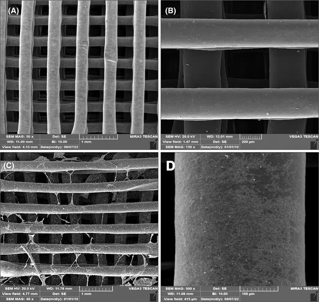
To tackle this challenge, a team of researchers designed a porous three-dimensional polymer scaffold using polycaprolactone, a biocompatible and flexible plastic filament widely used in medical prototyping. While the 3D printing process successfully creates a precise grid of square pores measuring approximately 350 micrometers across, the raw plastic surface remains too hydrophobic, meaning water and proteins slide right off instead of sticking around to encourage tissue growth. The work is published in FEBS Open Bio, detailing a multi-step chemical modification designed to transform the material interface.
How Chemical Etching and Silk Protein Coatings Transform Plastic Surfaces
To make the plastic friendly to living tissue, the research team first immersed the printed constructs in a sodium hydroxide solution for 24 hours. This chemical bath triggers hydrolysis on the outer surface of the plastic filaments, generating active hydroxyl groups that instantly improve surface wettability and lower the water contact angle from about 73 degrees down to 49 degrees.
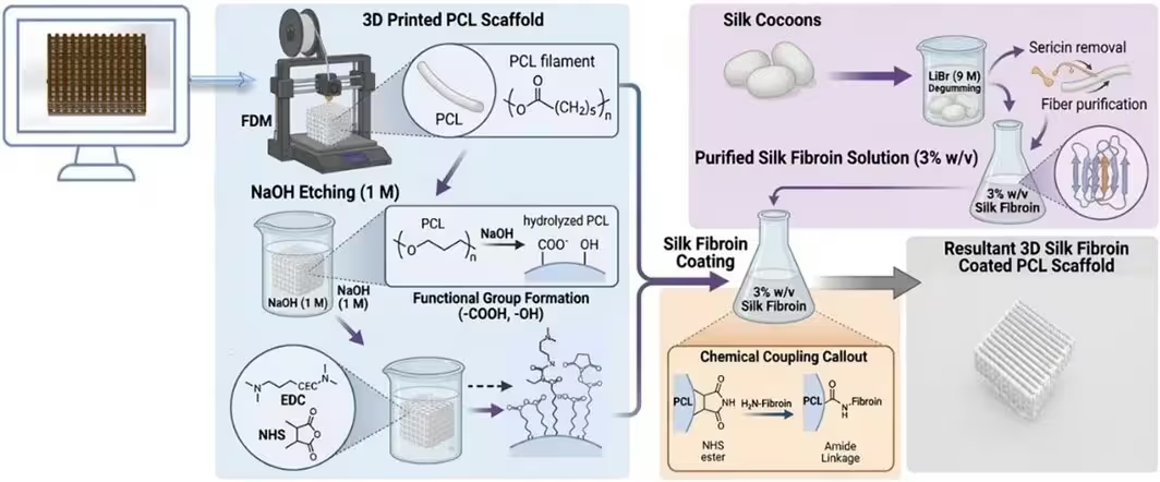
Next, the investigators applied a conformal coating of silk fibroin extracted from Bombyx mori silkworm cocoons. Using chemical cross-linking agents known as EDC and NHS, they permanently bonded the natural silk proteins to the etched plastic framework. Although the resulting water contact angle settled slightly higher at 52 degrees, the modified scaffolds retained high wettability while gaining complex biological signaling domains from the silk protein layer.
What the Laboratory Tests Revealed About Cell Growth and Safety
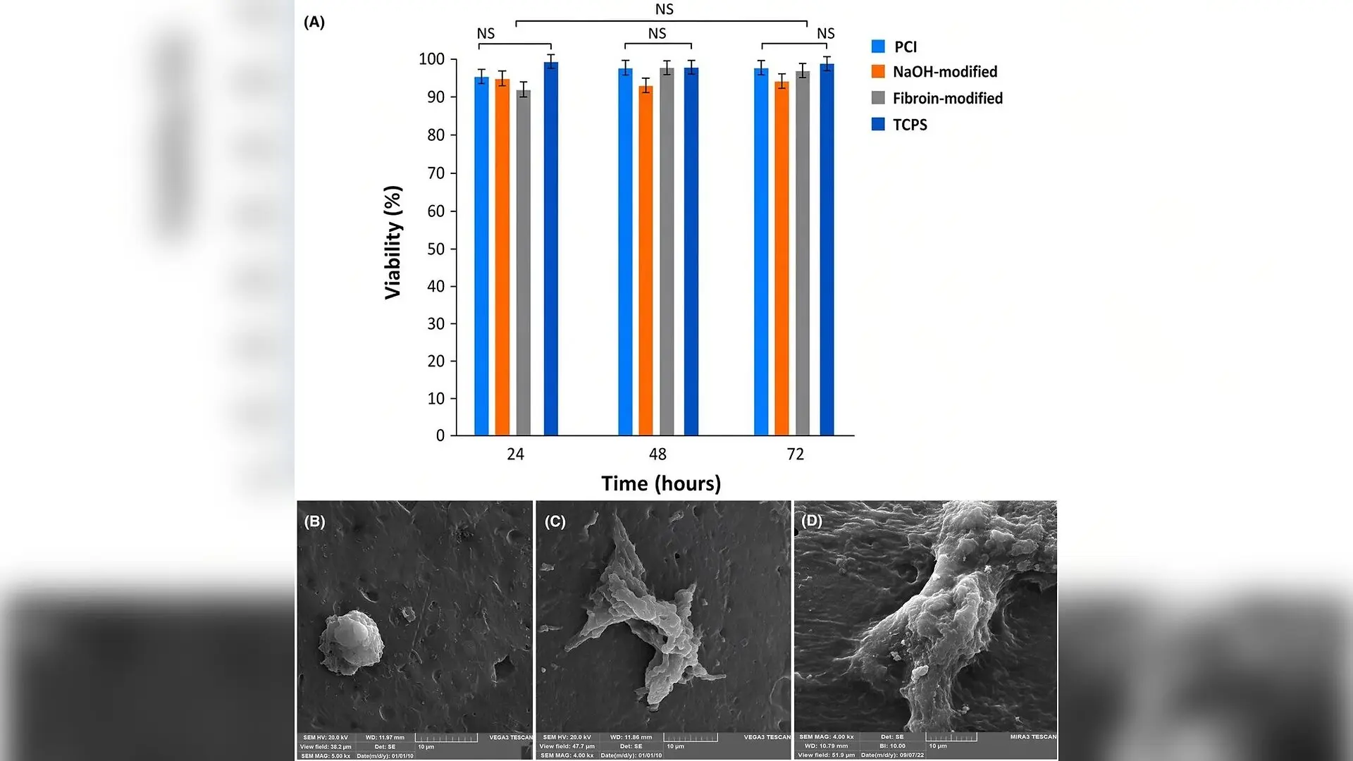
To see how human cells interact with the modified material, the team cultured human osteosarcoma-derived osteoblast-like cells directly on the scaffold disks. Over a 72-hour period, an MTT cell viability assay confirmed that the silk-coated constructs exhibited zero cytotoxicity, supporting normal metabolic activity and cell proliferation just as effectively as standard laboratory culture plates.
Scanning electron microscopy images provided a clear picture of cell morphology following the incubation period. While unmodified plastic scaffolds showed sparse cell attachment and poor spreading, the silk fibroin-coated surfaces supported a significantly higher density of well-spread cells anchored securely across the porous architecture.
What Limits Remain Before Clinical Application Can Begin?
Despite the promising laboratory results, the authors emphasize that these experiments represent an early in vitro phase of testing. The investigation did not evaluate long-term degradation kinetics, mechanical performance under heavy load-bearing conditions, or behavior inside dynamic fluid perfusion systems.
Future studies will need to test how the composite scaffolds perform in animal models with critical-sized bone defects to measure actual vascularization, new bone formation, and osseointegration. Additionally, researchers plan to study osteoprogenitor cell populations to confirm the upregulation of specific bone-forming markers like alkaline phosphatase and RUNX2 before moving toward eventual clinical translation.
The research was published in FEBS open bio on November 9, 2026.
This article has been fact checked for accuracy, with information verified against reputable sources. Learn more about us and our editorial process.
Last reviewed on .
Article history
- Latest version
Reference(s)
- Koeini, Hamed., et al. “Surface modification of
3D ‐printed polycaprolactone scaffolds with silk fibroin for bone tissue engineering.” FEBS Open Bio, November 9, 2026 Wiley, doi: 10.1002/2211-5463.70366. <https://doi.org/10.1002/2211-5463.70366>.
Cite this page:
- Posted by Robert Brown