New Titanium Pelvic Plate Shows Promise in Repairing Complex Dog Fractures
An anatomically contoured locking plate designed specifically for canine pelvises achieved reliable healing and preserved normal canal dimensions in a small case series.
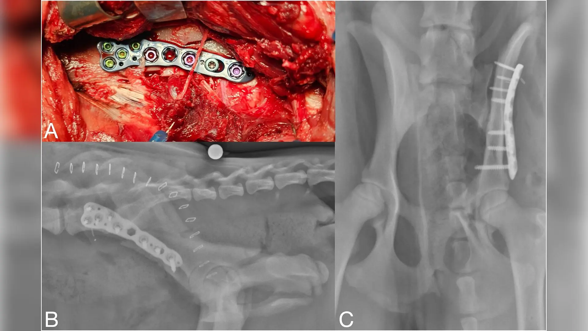
A custom-designed orthopedic plate made from titanium alloy has been evaluated for the surgical repair of broken hip bones in dogs, pointing to a reliable healing process and a low rate of complications in a small group of animal patients. Pelvic injuries represent up to a quarter of all fractures in cats and 16 percent in dogs, with motor vehicle accidents and high falls serving as the leading causes.
Why Traditional Pelvic Fixation Plates Fall Short
Conventional metal plates used to bridge breaks in the main pelvic bone, known as the ilial body, are rarely designed specifically for that anatomy. Surgeons typically rely on straight or reconstruction plates that require manual bending during operations and carry a roughly twentyfold higher risk of implant failure compared to locking systems. Because the pelvis experiences complex biomechanical forces surrounded by active muscle groups, inadequate fixation strength can lead to implant loosening, nonunion of the bone, or plate breakage.
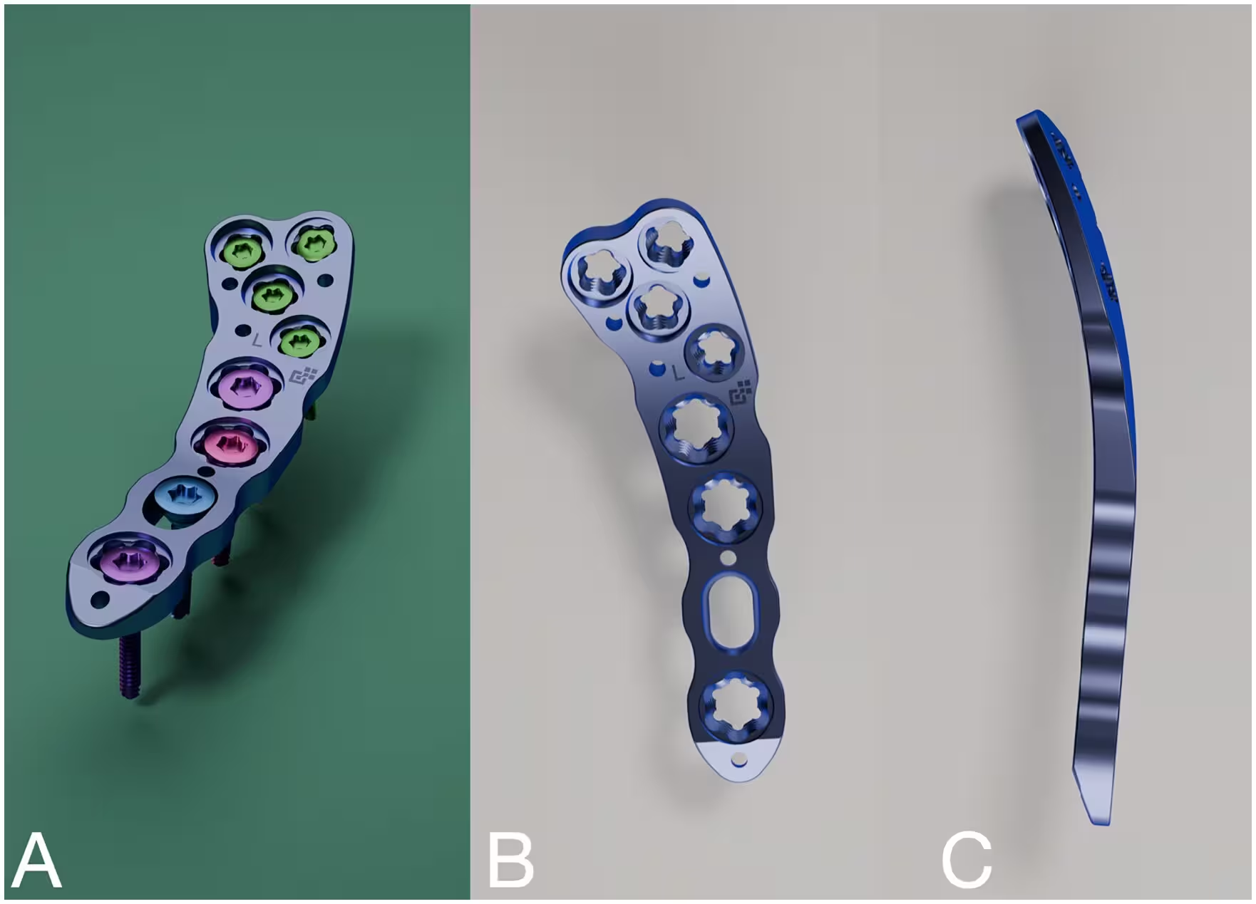
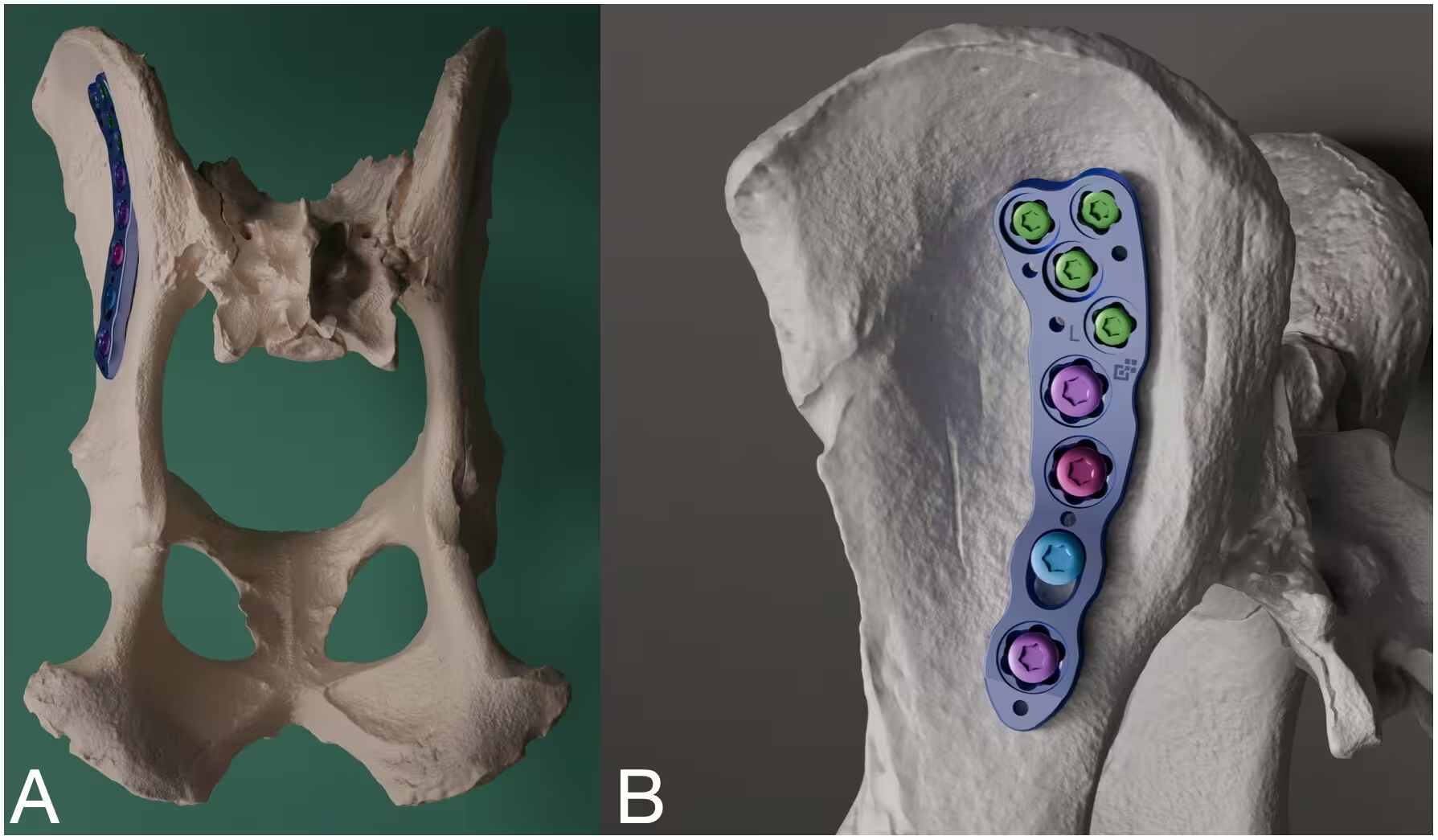
To address this anatomical mismatch, researchers developed a specialized hybrid implant called the 3.5 mm Steelvet Iliac Locking Plate. The engineering process utilized statistical shape modeling based on computed tomography scans from dogs weighing between 25 and 30 kilograms to map the natural curvature of the ilium. The resulting plate features side-specific contours, a wider proximal head to accommodate multiple locking screws in the thin bone of the iliac wing, and a narrow shaft designed to span the fracture toward the hip socket without requiring intraoperative bending.
Clinical Performance and Healing Outcomes in Ten Dogs
The retrospective investigation reviewed medical records and follow-up radiographs for ten client-owned dogs treated between 2023 and 2024. The study is published in PLOS One, detailing outcomes for animals ranging from a nine-month-old mixed-breed dog to a ten-year-old canine with weights spanning 21 to 52 kilograms. Breeds included German Shepherds, Great Pyrenees, Boxers, and Labrador Retrievers.
According to the findings, six of the ten dogs achieved complete radiographic bone union by 60 days postoperatively, and all ten dogs showed successful healing within 97 days. The median healing time settled at 60 days. Postoperative evaluations confirmed that pelvic canal dimensions remained within normal limits for every animal, preventing significant narrowing that could otherwise cause long-term complications like nerve compression or chronic constipation.
Understanding the Complications and Technical Limits
While the overall recovery trajectory proved favorable, one dog developed a localized cutaneous fistula, which is an abnormal skin opening near the surgical site, three months after the operation. Investigators noted no clinical or radiographic signs of deep bone infection, and the fracture itself was fully healed at the time the plate was surgically removed. The skin lesion resolved completely within seven days following the removal of the implant.
Despite the encouraging clinical observations, the authors emphasize that the study carries notable limitations. The retrospective design involved a small sample size of only ten animals, lacked a control group treated with standard plates, and did not incorporate objective functional metrics like force-plate gait analysis. Consequently, the findings demonstrate feasibility and safety rather than proven superiority over existing veterinary implants, leaving the door open for larger prospective trials to confirm the preliminary advantages.
The research was published in PLOS One on November 10, 2026.
This article has been fact checked for accuracy, with information verified against reputable sources. Learn more about us and our editorial process.
Last reviewed on .
Article history
- Latest version
Reference(s)
- Ferrigno, Cassio Ricardo Auada., et al. “Stabilization of canine ilial body fractures using an anatomically contoured locking iliac plate: A short retrospective case series.” PLOS One, vol. 21, no. 11, November 10, 2026, pp. e0352364 Public Library of Science (PLoS), doi: 10.1371/journal.pone.0352364. <https://doi.org/10.1371/journal.pone.0352364>.
Cite this page:
- Posted by Rohan Kumar