Updated Liver Cancer Imaging Criteria Resolve Post-Treatment Confusions
Recent guidelines refine how radiologists differentiate viable tumors from scar tissue and inflammation following complex therapies.
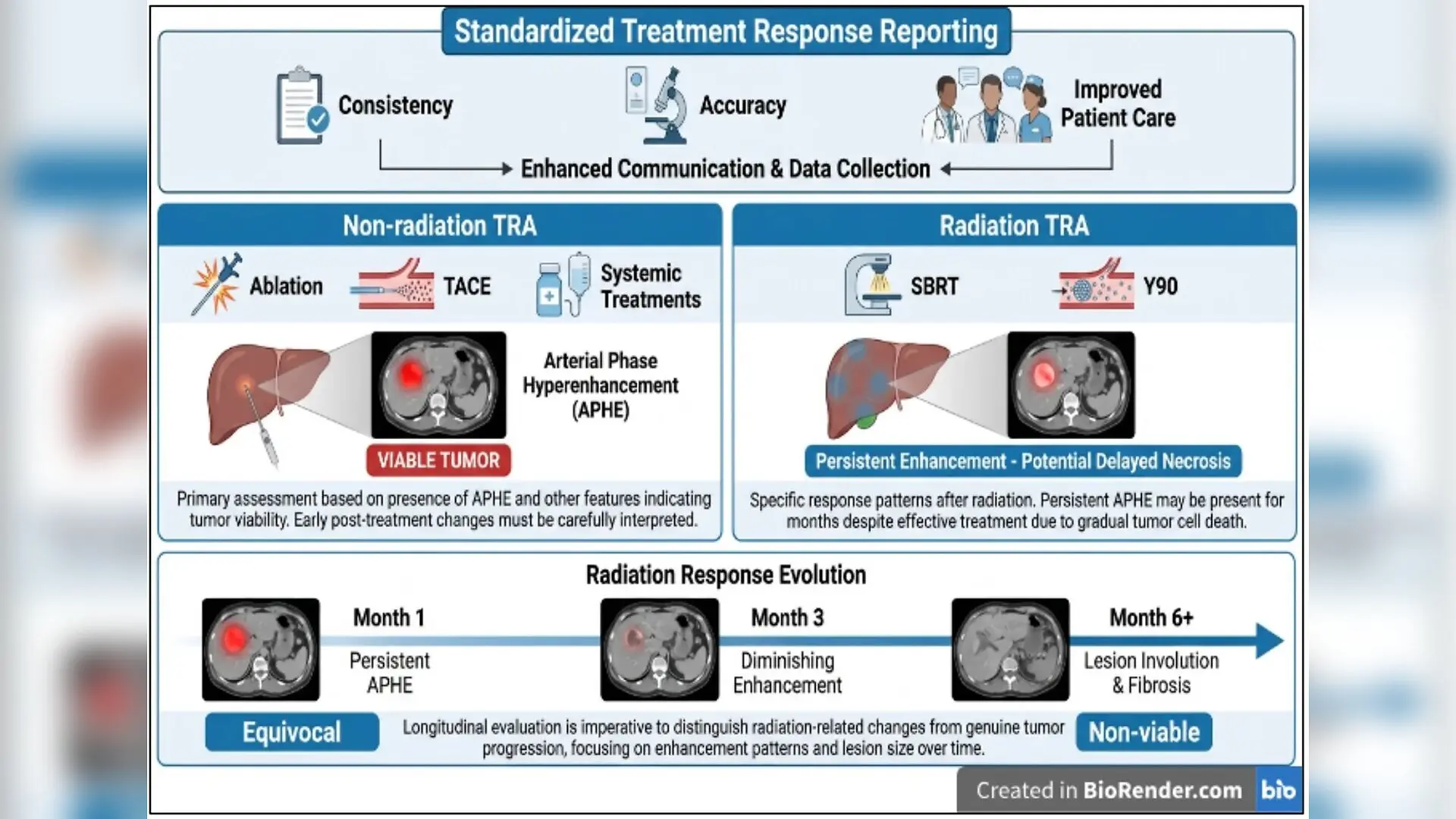
A new comprehensive review maps out critical updates to standard medical imaging criteria used to evaluate liver cancer treatments, addressing persistent diagnostic confusions like scar tissue and inflammation that mimic tumor growth. The detailed narrative evaluation, published in Cancer Management and Research in 2026, details how the updated Liver Imaging Reporting and Data System Treatment Response Algorithm (known as LI-RADS TRA) aims to bring much-needed consistency to oncology clinics worldwide.
How Modern Liver Cancer Treatments Changed Medical Imaging
Managing hepatocellular carcinoma, the most common type of primary liver cancer, has shifted dramatically over the past decade away from simple surgery toward complex multimodal strategies. Modern therapies now include thermal ablation, transarterial chemoembolization (which involves blocking blood flow while delivering localized chemotherapy), stereotactic body radiotherapy, and immune checkpoint inhibitors. While these advanced approaches extend patient survival, they also create highly complex post-treatment imaging appearances that often confuse radiologists.
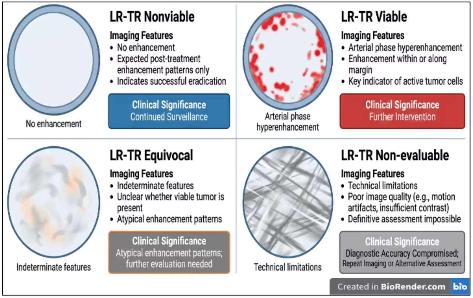
Traditional response criteria such as RECIST rely strictly on measuring tumor shrinkage. However, many successful locoregional therapies kill cancer cells without shrinking the overall mass immediately, leaving behind dead tissue or scarring that can look deceptively like active disease on standard scans. The LI-RADS framework was built specifically to solve this problem by focusing on lesion-level viability rather than total size, classifying treated areas based on specific vascular and enhancement patterns.
What the Updated 2024-2025 Framework Changes for Clinicians
Led by researchers Arnabjyoti Deva Sarma, Debanga Sarma, and Thomas J Webster, the review outlines major structural refinements introduced in the v2024-2025 guidelines. Most notably, the updated algorithm separates non-radiation therapies from radiation-based treatments into two distinct evaluation pathways. Radiation treatments like stereotactic body radiotherapy and yttrium-90 radioembolization frequently cause persistent arterial phase hyperenhancement for months even when the treatment is completely successful, due to delayed radiation-induced necrosis.
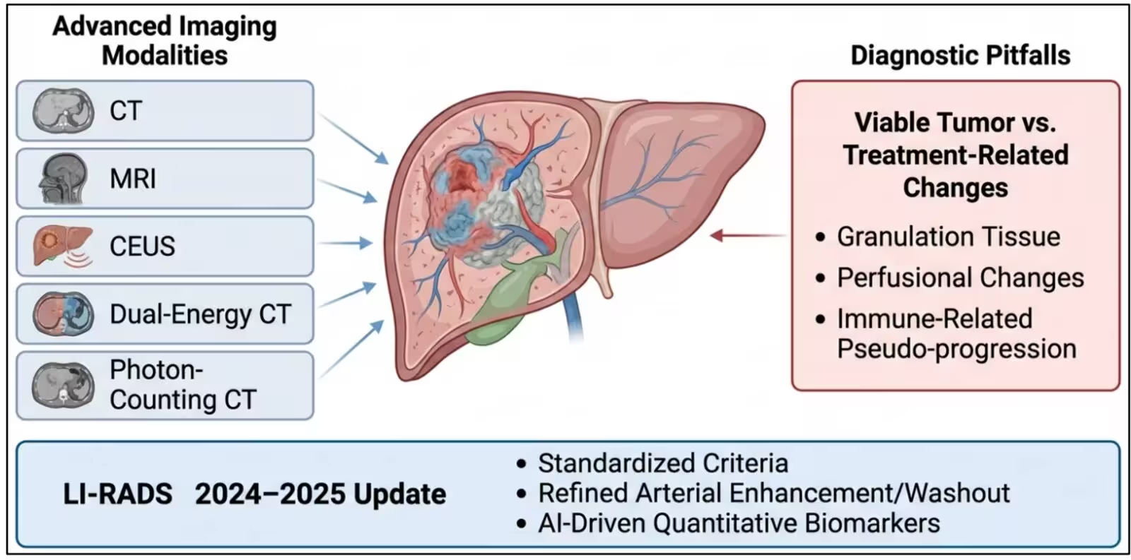
The revised guidelines also provide clearer instructions for interpreting major features like arterial phase hyperenhancement and contrast washout, alongside ancillary features like diffusion restriction on magnetic resonance imaging. By integrating multiparametric magnetic resonance imaging and advanced contrast-enhanced ultrasound, clinicians can better differentiate between active, viable tumor cells and benign post-procedural changes such as granulation tissue, regional hyperemia, or fat deposition.
Navigating Immunotherapy Pitfalls and Artificial Intelligence Horizons
One of the most pressing challenges highlighted in the review is evaluating patients receiving systemic immunotherapies. These modern agents can trigger immune cell infiltration and localized inflammation that temporarily enlarges the tumor or creates new lesions before shrinking them, a phenomenon known as pseudoprogression. Misinterpreting this immune flare as true treatment failure can lead doctors to halt effective therapy prematurely. To combat this, the review points to specialized immune-related criteria like iRECIST as essential additions to modern diagnostic workflows.
Looking toward the future, the authors emphasize the growing potential of artificial intelligence, radiomics, and deep learning models to automate complex image interpretation. By extracting high-dimensional quantitative features from medical scans that remain invisible to the human eye, computational algorithms could soon help predict treatment resistance and recurrence risk long before traditional volumetric changes appear. Nevertheless, the researchers caution that these advanced tools remain largely confined to single-center retrospective studies, requiring rigorous multi-center prospective validation before becoming standard clinical practice.
The research was published in Cancer management and research on January 1, 2026.
This article has been fact checked for accuracy, with information verified against reputable sources. Learn more about us and our editorial process.
Last reviewed on .
Article history
- Latest version
Reference(s)
- Sarma, Arnabjyoti Deva., et al. “Advances in the LI-RADS Treatment Response Algorithm: Imaging Principles, Diagnostic Challenges, and Clinical Integration Following the LI-RADS 2024 Update.” Cancer Management and Research, vol. Volume 18, October 1, 2026, pp. 1-29. Informa UK Limited, doi: 10.2147/CMAR.S614483. <https://doi.org/10.2147/CMAR.S614483>.
Cite this page:
- Posted by Robert Brown