Genetically Modified Rodent Parasites Reveal Clues for Vivax Malaria Vaccine
Biotechnology

Genetically Modified Rodent Parasites Reveal Clues for Vivax Malaria Vaccine

Scientists engineered surrogate malaria parasites carrying human-infecting P. vivax antigens, achieving partial lab inhibition and limited living protection in mice.
By Robert Brown
Published:
Email this Article
Genetically Modified Rodent Parasites Reveal Clues for Vivax Malaria Vaccine
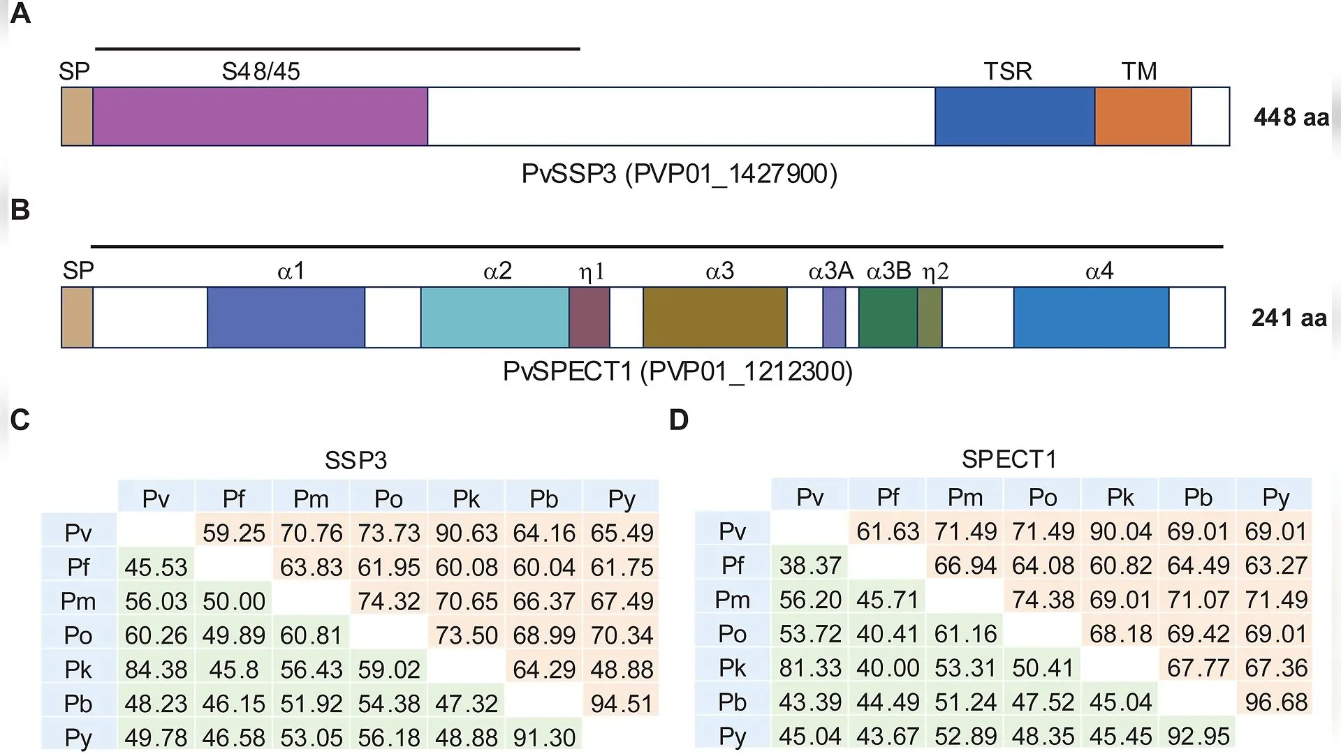
Features of antigens selected. (A) Schematic representation of P. vivax SSP3 protein showing different domains. SP - signal peptide, S48/45 - S48/45 domain of Plasmodium 6-Cys proteins, TSR - thrombospondin repeat, TM - transmembrane domain. The top line represents the region (AA 19-203) selected for recombinant PvSSP3 production. (B) Schematic representation of P. vivax SPECT1 protein showing different domains. SP - signal peptide, α1, α2, α3 and α4 - four different alpha helices in the bundle, α3a and α3b - antiparallel α-helices in the hook structure. The black line at the top represents the region selected for recombinant PvSPECT1 production. (C and D) Homology and Identity matrices of SSP3 and SPECT1 from different Plasmodium species were calculated using the sequence manipulation suite [61] (C) Homology and identity matrix of SSP3. Pv - P. vivax P01 (PVP01_1427900), Pf - P. falciparum 3D7 (PF3D7_0812300), Pm - P. malariae UG01 (PmUG01_14043900), Po - P. ovale curtisi GH01 (PocGH0 Surendra Kumar Kolli et al. / PLOS Neglected Tropical Diseases

Finding new ways to block malaria before it infects human liver cells remains a major global health hurdle, especially because the parasite Plasmodium vivax creates dormant liver stages that trigger relapses months or years later. Researchers have long struggled to test human vaccine candidates because obtaining enough laboratory-produced sporozoites, the infectious stage injected by mosquitoes, is extremely difficult without human or primate hosts.

To bypass this bottleneck, a research team engineered surrogate rodent malaria parasites carrying two key human vaccine targets. The study, published in PLOS Neglected Tropical Diseases, details how scientists used a genetic replacement strategy to swap native parasite genes in Plasmodium berghei with their human-infecting P. vivax counterparts, creating stable chimeric lines for laboratory evaluation.

How Rodent Parasites Became Stand-Ins for Human Malaria Targets

The research team focused on two specific pre-erythrocytic proteins known as SSP3, standing for sporozoite surface protein 3, and SPECT1, standing for sporozoite microneme protein essential for cell traversal. Both proteins play vital roles when the parasite moves through skin tissue, crosses liver barriers, and invades host cells. Using a two-step gene insertion and marker-removal technique, the authors successfully replaced the mouse parasite genes with the full-length coding sequences for P. vivax SSP3 and SPECT1.
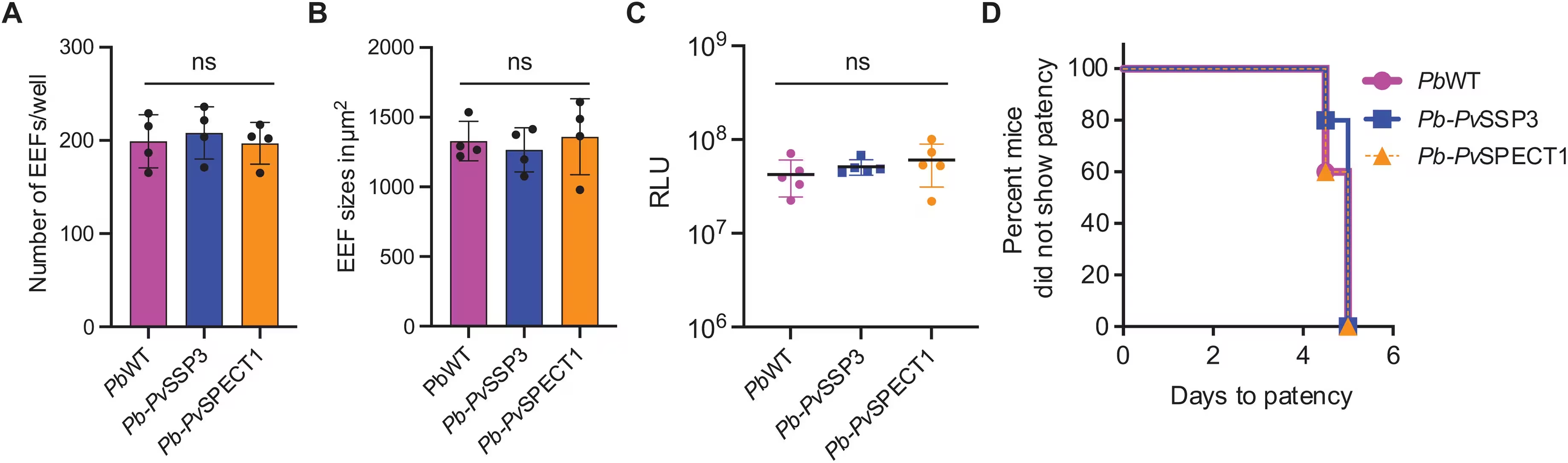
Fig 5: In vitro and in vivo infectivity of Pb-PvSSP3 and Pb-PvSPECT1 sporozoites. (A) Bar graph showing the number of EEFs per well at 48 hours post infection (hpi) in an HC-04 monolayer infected with 2 x 10⁴ of PbWT, Pb-PvSSP3 or Pb-PvSPECT1 sporozoites in a 384-well plate. Data were obtained from 4 independent experiments, each with at least 8 wells. Error bars represent the mean with the standard deviation. A Shapiro-Wilk test confirmed that the EEF numbers/well were normally distributed. Significance was calculated using One-way ANOVA with Tukey’s multiple comparisons test (p = 0.88 for Pb-PvSSP3 and p = 0.99 for Pb-PvSPECT1), ns - not significant. (B) Bar graph showing the average size of EEF in µm2 at 48 hpi in an HC-04 monolayer infected with PbWT, Pb-PvSSP3 or Pb-PvSPECT1 sporozoites in a 384-well plate. Data are obtained from 4 independent experiments, each with at least 1000 EEFs. Error bars represent the mean with the standard deviation. A Shapiro-Wilk test confirmed that the EEF s
In vitro and in vivo infectivity of Pb-PvSSP3 and Pb-PvSPECT1 sporozoites. (A) Bar graph showing the number of EEFs per well at 48 hours post infection (hpi) in an HC-04 monolayer infected with 2 x 10⁴ of PbWT, Pb-PvSSP3 or Pb-PvSPECT1 sporozoites in a 384-well plate. Data were obtained from 4 independent experiments, each with at least 8 wells. Error bars represent the mean with the standard deviation. A Shapiro-Wilk test confirmed that the EEF numbers/well were normally distributed. Significance was calculated using One-way ANOVA with Tukey’s multiple comparisons test (p = 0.88 for Pb-PvSSP3 and p = 0.99 for Pb-PvSPECT1), ns - not significant. (B) Bar graph showing the average size of EEF in µm2 at 48 hpi in an HC-04 monolayer infected with PbWT, Pb-PvSSP3 or Pb-PvSPECT1 sporozoites in a 384-well plate. Data are obtained from 4 independent experiments, each with at least 1000 EEFs. Error bars represent the mean with the standard deviation. A Shapiro-Wilk test confirmed that the EEF s Credit: Surendra Kumar Kolli et al. / PLOS Neglected Tropical Diseases

A series of biological fitness tests confirmed that these genetic modifications did not harm the parasite. The transgenic organisms developed normally in both mice and mosquitoes, producing oocysts and salivary gland sporozoites in numbers comparable to wild-type strains. When researchers introduced these modified sporozoites into human hepatoma cell monolayers, the parasites demonstrated typical growth and liver-stage development.

Antibody Inhibition and In Vivo Challenge Results

After confirming that the transgenic parasites matured normally, the investigators tested whether antibodies raised against recombinant versions of SSP3 and SPECT1 could block infection. In laboratory assays, antisera directed against PvSSP3 reduced hepatocyte invasion by a mean of 27 percent, while antisera against PvSPECT1 achieved a mean inhibition of 14 percent.

However, the transition from laboratory inhibition to living animal protection yielded mixed outcomes. When vaccinated mice were challenged with the transgenic parasites via intravenous injection, animals immunized with the PvSSP3 candidate showed no statistically significant protection compared to control groups. In contrast, mice immunized with the PvSPECT1 candidate achieved sterile protection in 10 percent of the test subjects, meaning those animals remained entirely free of blood-stage parasitemia.
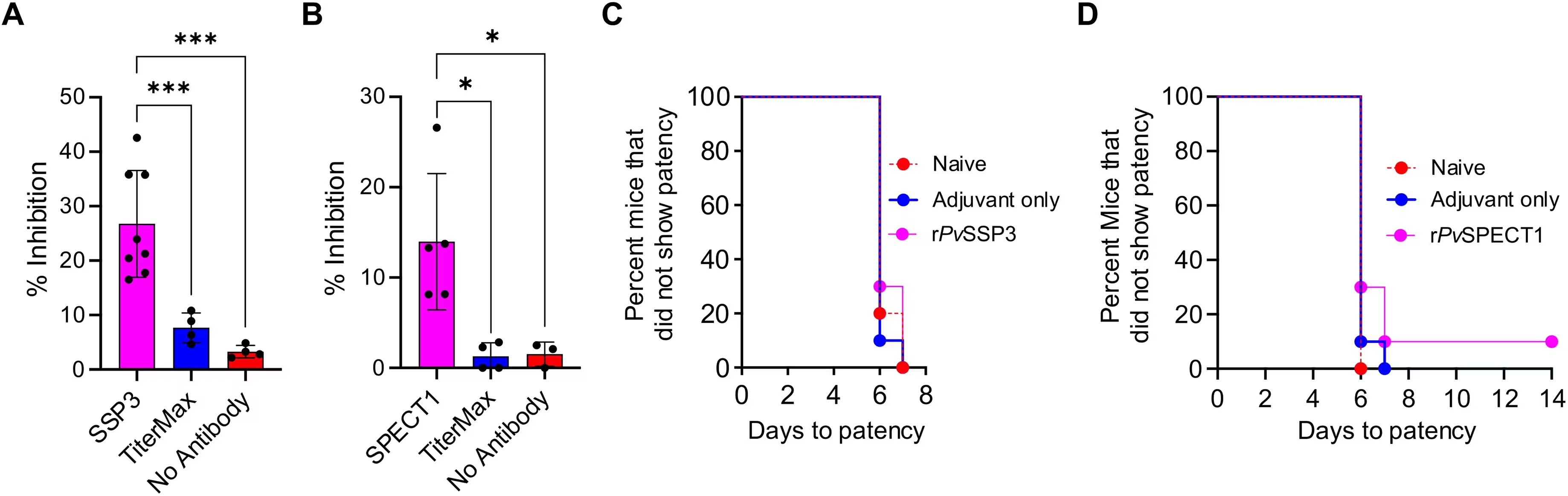
Fig 6: Antisera against PvSSP3 and PvSPECT1 inhibit hepatocyte invasion, and PvSPECT1 confers limited protection in BALB/c mice after sporozoite challenge. (A and B) In vitro inhibition of liver-stage development assay (ILSDA) using 2 x 10³ transgenic P. berghei sporozoites expressing PvSSP3 (Pb-PvSSP3) or PvSPECT1 (Pb-PvSPECT1). Immune sera were generated by immunizing BALB/c mice with recombinant PvSSP3 or PvSPECT1 adjuvanted with Titremax Gold [7]. Pb-PvSSP3 or Pb-PvSPECT1 sporozoites were incubated with 1:100 dilution of the immune sera for 20 min at room temperature and added to monolayers of HC-04 line in 384-well plates for infection. Reduction in infection was expressed as a percentage inhibition of EEF formation by the immune sera relative to pre-immune serum. (A) Bar graph showing the percent inhibition of Pb-PvSSP3 EEFs by PvSSP3 immune serum. Each dot represents the serum from an individual mouse (n = 8), and the data were obtained from 4 independent experiments, each containing e
Antisera against PvSSP3 and PvSPECT1 inhibit hepatocyte invasion, and PvSPECT1 confers limited protection in BALB/c mice after sporozoite challenge. (A and B) In vitro inhibition of liver-stage development assay (ILSDA) using 2 x 10³ transgenic P. berghei sporozoites expressing PvSSP3 (Pb-PvSSP3) or PvSPECT1 (Pb-PvSPECT1). Immune sera were generated by immunizing BALB/c mice with recombinant PvSSP3 or PvSPECT1 adjuvanted with Titremax Gold [7]. Pb-PvSSP3 or Pb-PvSPECT1 sporozoites were incubated with 1:100 dilution of the immune sera for 20 min at room temperature and added to monolayers of HC-04 line in 384-well plates for infection. Reduction in infection was expressed as a percentage inhibition of EEF formation by the immune sera relative to pre-immune serum. (A) Bar graph showing the percent inhibition of Pb-PvSSP3 EEFs by PvSSP3 immune serum. Each dot represents the serum from an individual mouse (n = 8), and the data were obtained from 4 independent experiments, each containing e Credit: Surendra Kumar Kolli et al. / PLOS Neglected Tropical Diseases

Why Laboratory Inhibition Might Not Equal Full Protection

The study authors note that achieving only modest in vivo protection despite successful antibody generation highlights a common hurdle in malaria vaccine development. Partial inhibition in glass dishes does not automatically translate into sterilizing immunity inside a living host organism. Furthermore, the recombinant construct used for SSP3 covered only a partial fragment of the protein, which may have lacked critical structural folding needed to elicit strong neutralizing antibodies.

Another crucial factor involves the method of infection. Injecting sporozoites directly into the bloodstream via intravenous routes bypasses the natural skin stage, a vital phase where antibodies normally act to restrict parasite motility and block vascular access. The authors suggest that future evaluations should test these vaccine candidates using natural mosquito bites or intradermal delivery routes, alongside more potent clinical adjuvants, to determine their true potential for eliminating P. vivax malaria.

The research was published in PLOS Neglected Tropical Diseases on October 22, 2026.

Fact Checked

This article has been fact checked for accuracy, with information verified against reputable sources. Learn more about us and our editorial process.

Last reviewed on .

Article history

  • Latest version

Reference(s)

  1. Kolli, Surendra Kumar., et al. “Transgenic Plasmodium berghei expressing P. vivax pre-erythrocytic antigens SSP3 and SPECT1 to elucidate their efficacy towards vaccine development.” PLOS Neglected Tropical Diseases, vol. 20, no. 10, October 22, 2026, pp. e0014810 Public Library of Science (PLoS), doi: 10.1371/journal.pntd.0014810. <https://doi.org/10.1371/journal.pntd.0014810>.

Cite this page:

Brown, Robert. “Genetically Modified Rodent Parasites Reveal Clues for Vivax Malaria Vaccine.” BioScience. BioScience ISSN 2521-5760, 27 October 2026. <https://www.bioscience.com.pk/en/subject/biotechnology/transgenic-plasmodium-berghei-expressing-p-vivax-pre-erythrocytic-antigens-ssp3-and-spect1-to-elucidate-their-efficacy-towards-vaccine-development>. Brown, R. (2026, October 27). “Genetically Modified Rodent Parasites Reveal Clues for Vivax Malaria Vaccine.” BioScience. ISSN 2521-5760. Retrieved October 27, 2026 from https://www.bioscience.com.pk/en/subject/biotechnology/transgenic-plasmodium-berghei-expressing-p-vivax-pre-erythrocytic-antigens-ssp3-and-spect1-to-elucidate-their-efficacy-towards-vaccine-development Brown, Robert. “Genetically Modified Rodent Parasites Reveal Clues for Vivax Malaria Vaccine.” BioScience. ISSN 2521-5760. https://www.bioscience.com.pk/en/subject/biotechnology/transgenic-plasmodium-berghei-expressing-p-vivax-pre-erythrocytic-antigens-ssp3-and-spect1-to-elucidate-their-efficacy-towards-vaccine-development (accessed October 27, 2026).
End of the article