Scientists Build Synthetic DNA Complexes to Solve a Bacterial Gene-Transfer Mystery
Biotechnology

Scientists Build Synthetic DNA Complexes to Solve a Bacterial Gene-Transfer Mystery

A newly developed in vitro system reveals how plant and animal enzymes clear protein roadblocks during bacterial DNA integration.
By Robert Brown
Published:
Email this Article
Scientists Build Synthetic DNA Complexes to Solve a Bacterial Gene-Transfer Mystery
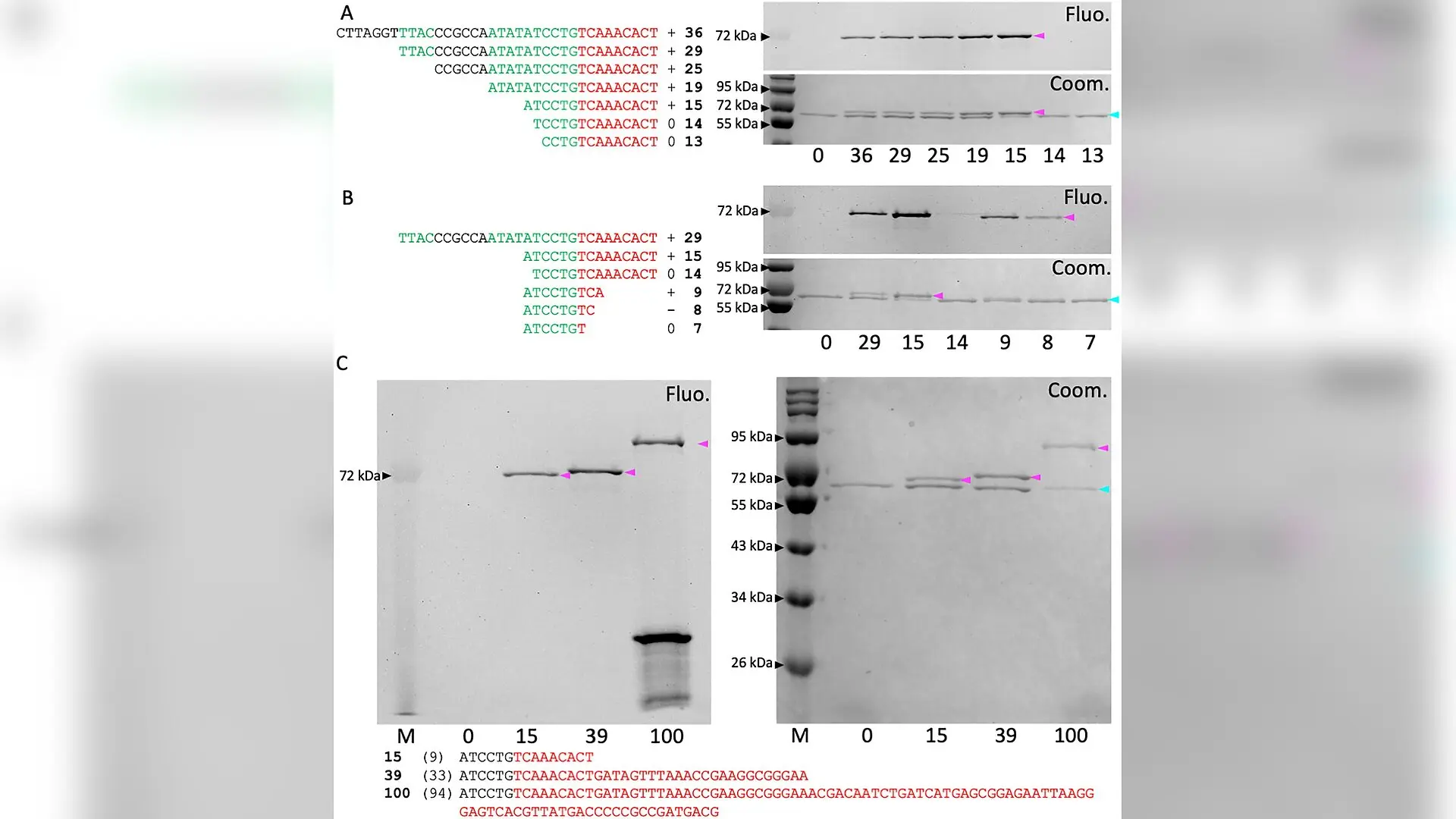
Synthetic T-complexes with different lengths of ssDNA. (A) Reducing the length of the ssDNA substrate from its 5’-end. “+”, “-“, and “0” indicate, respectively, efficient, weak, or absent formation of the VirD2-ssDNA complex. (B) Reducing the length of the ssDNA substrate from its 3’-end. Each lane is labeled with the total number of nt in the ssDNA substrate; the two highly conserved sequences within the 25-bp of the right border are indicated in green; sequences that remained attached to VirD2 in the VirD2-ssDNA complex are indicated in red. (C) Increasing the length of the ssDNA substrate. Each lane is labeled with the total number of nt in the ssDNA substrate; numbers of nt that remain attached to VirD2 are in parentheses; VirD2 and VirD2-ssDNA complexes are indicated by cyan and magenta arrowheads, respectively. Benoît Lacroix et al. / PLOS Pathogens

Scientists have developed a cell-free technique to track how a plant-infecting bacterium hands off its genetic material, revealing a key biochemical checkpoint that could eventually refine plant genetic engineering methods.

How Agrobacterium Transfers DNA Into Host Genomes

The bacterium Agrobacterium tumefaciens causes crown gall disease in plants by slipping a segment of its plasmid DNA, known as T-DNA, directly into the host cell genome. Researchers have long used this natural gene-transfer system as a primary tool for modifying plant and fungal genomes in laboratories worldwide. However, the exact molecular steps governing how this foreign DNA successfully integrates into host chromosomes remain incompletely understood.

During the infection process, the bacterial protein VirD2 acts as a specialized endonuclease, an enzyme that cuts DNA strands. VirD2 cleaves the bacterial plasmid at specific boundary sequences and stays covalently bound to the resulting single-stranded DNA segment, forming a nucleoprotein unit called the T-complex. This attached protein acts as a physical roadblock that prevents host enzymes from sealing the DNA ends into the plant genome. To complete integration, the VirD2 protein must be cleaved and removed.

Building Synthetic T-Complexes in the Laboratory

To study this removal process in detail, researchers designed a simplified in vitro system to generate artificial T-complexes. By incubating purified wild-type VirD2 protein with synthetic single-stranded DNA fragments tagged with a fluorescent dye at one end, the team successfully reconstituted the protein-DNA complexes outside of living cells. The findings were published in PLOS Pathogens in October 2026 by molecular biologists Benoît Lacroix and Vitaly Citovsky.
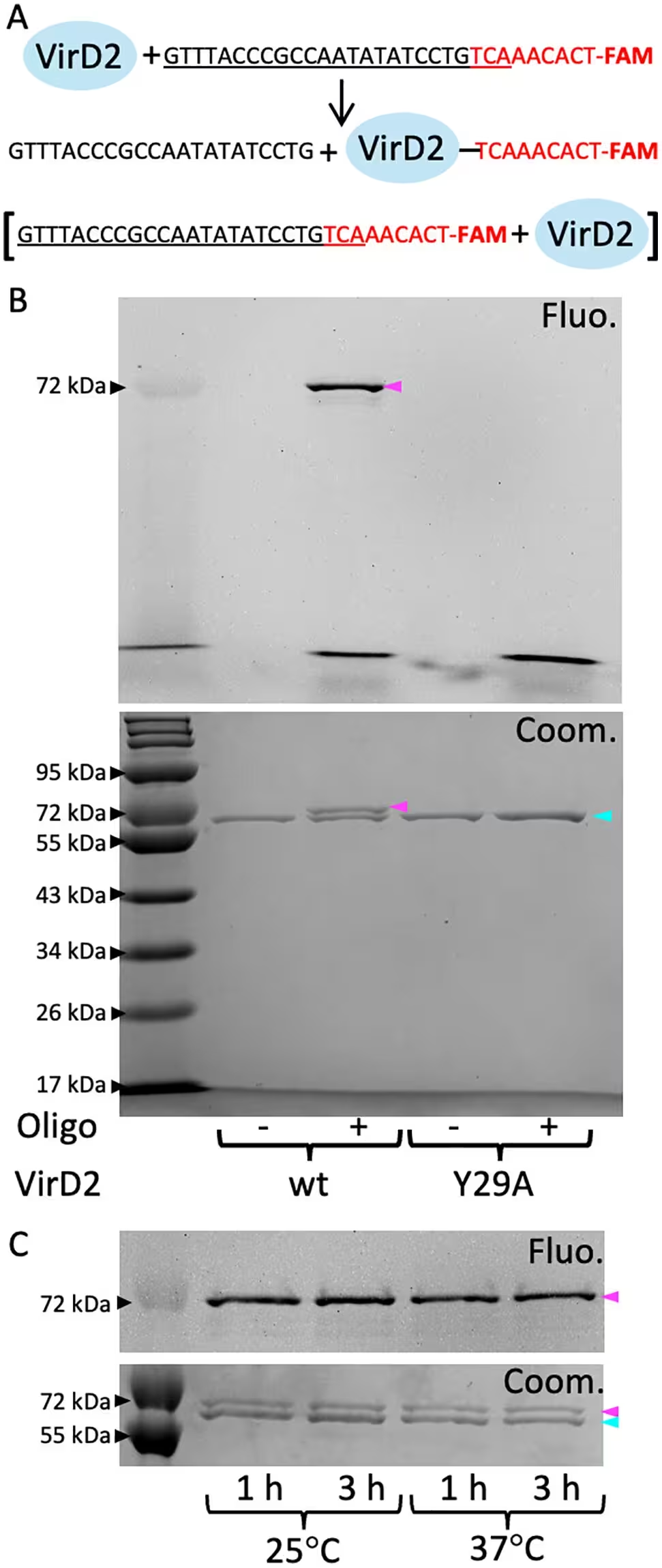
Fig 1: Generation of fluorescently labeled VirD2-ssDNA complexes. (A) Schematic illustration of the reaction between VirD2 and a ssDNA substrate. Incubation of purified VirD2 and an ssDNA oligomer containing the T-DNA right border (RB) sequence of the Ti-plasmid of the A. tumefaciens C58 strain and fluorescently tagged with FAM results in cleavage at RB and generation of a VirD2-ssDNA complex. The 25-bp sequence corresponding to the T-DNA right border (RB) is underlined, and sequences that remain attached to VirD2 in the VirD2-ssDNA complex are indicated in red. (B) Electrophoretic analysis of VirD2-ssDNA complex formation. The reaction mixtures were resolved by SDS-PAGE on a 10% gel, and free VirD2 and VirD2-ssDNA complexes were visualized by fluorescence imaging (Fluo., top panel) and Coomassie blue staining (Coom., bottom panel). (C) Effects of temperature and incubation time on VirD2-ssDNA complex formation. The 72-kDa size marker is indicated by a black arrowhead; VirD2 and VirD2-ssDNA c
Generation of fluorescently labeled VirD2-ssDNA complexes. (A) Schematic illustration of the reaction between VirD2 and a ssDNA substrate. Incubation of purified VirD2 and an ssDNA oligomer containing the T-DNA right border (RB) sequence of the Ti-plasmid of the A. tumefaciens C58 strain and fluorescently tagged with FAM results in cleavage at RB and generation of a VirD2-ssDNA complex. The 25-bp sequence corresponding to the T-DNA right border (RB) is underlined, and sequences that remain attached to VirD2 in the VirD2-ssDNA complex are indicated in red. (B) Electrophoretic analysis of VirD2-ssDNA complex formation. The reaction mixtures were resolved by SDS-PAGE on a 10% gel, and free VirD2 and VirD2-ssDNA complexes were visualized by fluorescence imaging (Fluo., top panel) and Coomassie blue staining (Coom., bottom panel). (C) Effects of temperature and incubation time on VirD2-ssDNA complex formation. The 72-kDa size marker is indicated by a black arrowhead; VirD2 and VirD2-ssDNA c Credit: Benoît Lacroix et al. / PLOS Pathogens

Through this system, the authors established that the tyrosine residue at position 29 of the VirD2 protein is strictly required for its DNA-nicking activity. When they substituted this tyrosine with an alanine residue, the resulting mutant protein completely lost its ability to bind and cleave the DNA strands. Testing various DNA lengths revealed that an eight-nucleotide sequence represents the minimal region required for VirD2 recognition and processing.

Clearing the Roadblock With Tyrosyl-DNA Phosphodiesterase 2

With a reliable in-laboratory model established, the team investigated whether tyrosyl-DNA phosphodiesterase 2 (TDP2), an enzyme known for repairing damaged DNA-protein crosslinks in eukaryotes, could sever VirD2 from the synthetic complexes. Using both commercially available human TDP2 and partially purified plant TDP2 from Arabidopsis thaliana, the researchers tracked the enzymatic cleavage of the artificial T-complexes.
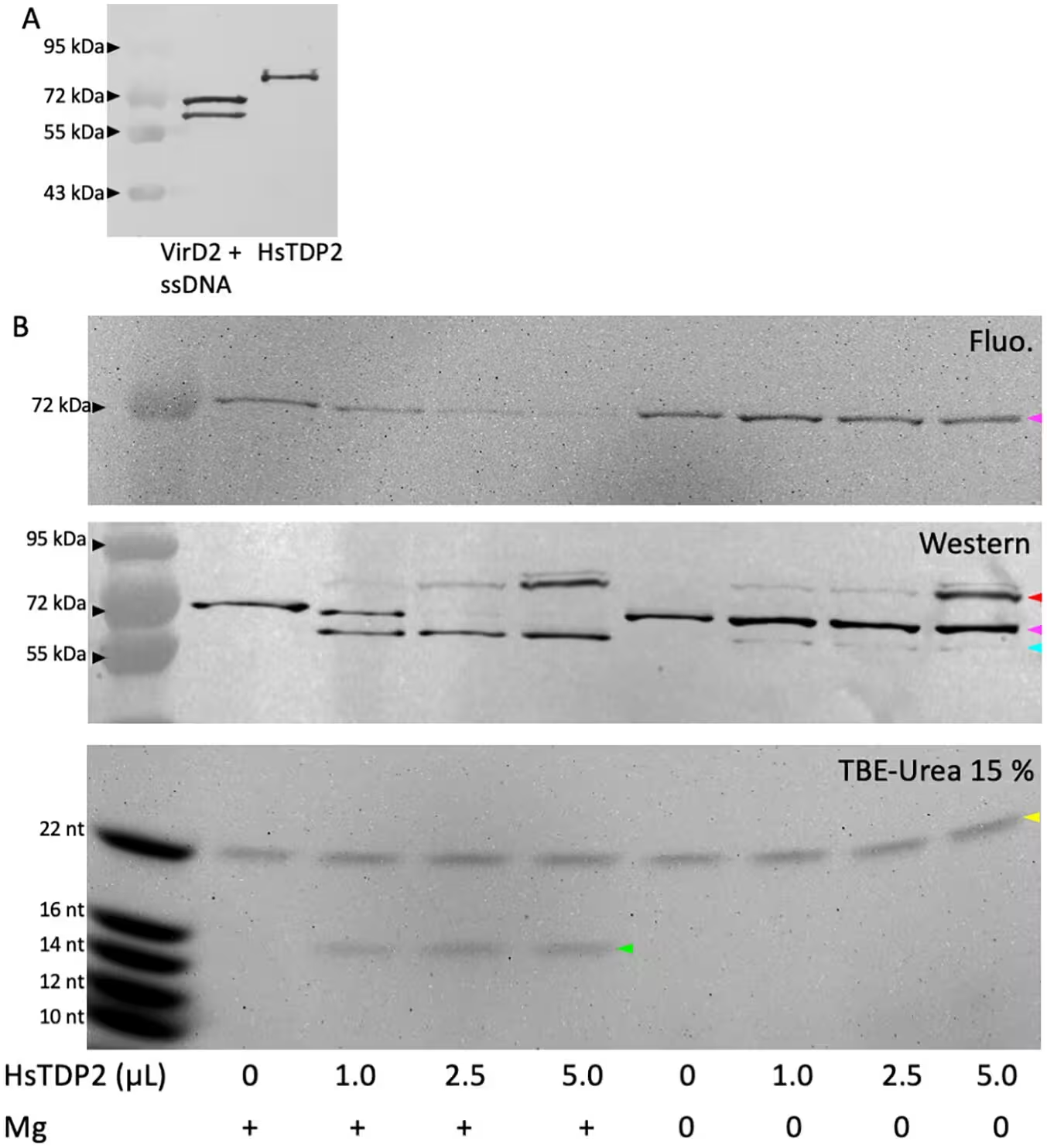
Fig 3: Removal of VirD2 from the synthetic T-complex by HsTDP2. (A) Western blot detection of VirD2-ssDNA and HsTDP2 using anti-6xHis antibodies. (B) Electrophoretic analysis of VirD2 removal by HsTDP2. VirD2-ssDNA complexes were incubated without HsTDP2 or with increasing concentrations of HsTDP2, in the presence or absence of MgCl2. The reaction mixtures were resolved by SDS-PAGE on a 10% gel, and free VirD2 and VirD2-ssDNA complexes were visualized by fluorescence imaging (Fluo., top panel) and Western blotting (Western, middle panel). Free VirD2, VirD2-ssDNA complexes, and free HsTDP2 are indicated by cyan, magenta, and red arrowheads, respectively. The sizes of ssDNA liberated by HsTDP2 were analyzed by electrophoresis on TBE-Urea 15% gels (TBE-Urea 15%, bottom panel). 16-nt and 22-nt ssDNA oligomers are indicated by green and yellow arrowheads, respectively.
Removal of VirD2 from the synthetic T-complex by HsTDP2. (A) Western blot detection of VirD2-ssDNA and HsTDP2 using anti-6xHis antibodies. (B) Electrophoretic analysis of VirD2 removal by HsTDP2. VirD2-ssDNA complexes were incubated without HsTDP2 or with increasing concentrations of HsTDP2, in the presence or absence of MgCl2. The reaction mixtures were resolved by SDS-PAGE on a 10% gel, and free VirD2 and VirD2-ssDNA complexes were visualized by fluorescence imaging (Fluo., top panel) and Western blotting (Western, middle panel). Free VirD2, VirD2-ssDNA complexes, and free HsTDP2 are indicated by cyan, magenta, and red arrowheads, respectively. The sizes of ssDNA liberated by HsTDP2 were analyzed by electrophoresis on TBE-Urea 15% gels (TBE-Urea 15%, bottom panel). 16-nt and 22-nt ssDNA oligomers are indicated by green and yellow arrowheads, respectively. Credit: Benoît Lacroix et al. / PLOS Pathogens

The biochemical assays showed that both animal and plant versions of TDP2 successfully removed the VirD2 protein from the single-stranded DNA segments. This activity required magnesium ions, matching the known dependence of mammalian TDP2 on divalent cations. When researchers introduced a specific point mutation into the plant enzyme, replacing a conserved histidine at position 431 with an asparagine, the enzyme lost its cleaving capability entirely. Furthermore, fluorescence and western blot analyses confirmed that TDP2 specifically hydrolyzed the bond between the VirD2 tyrosine residue and the first nucleotide of the DNA without damaging the rest of the strand.

Broader Context and What Lies Ahead

These findings provide direct biochemical evidence supporting the hypothesis that host TDP2 serves as a clearance mechanism during bacterial genetic transformation. Because both human and plant versions of the enzyme successfully processed the artificial complexes, the mechanism appears to be evolutionarily conserved, which aligns with the known ability of Agrobacterium to transfer genes into a wide array of eukaryotic species.

While genetic studies have previously indicated that TDP2 mutations reduce transformation efficiency, this work offers a direct view of the protein-cleaving events required for successful genomic integration. Future investigations utilizing mutated versions of VirD2 and various host factors will help clarify how these molecular interactions influence stable genetic transformations in biotechnology applications.

The research was published in PLOS Pathogens on October 26, 2026.

Fact Checked

This article has been fact checked for accuracy, with information verified against reputable sources. Learn more about us and our editorial process.

Last reviewed on .

Article history

  • Latest version

Reference(s)

  1. Lacroix, Benoît., et al. “Removal of VirD2 from a synthetic T-DNA complex of Agrobacterium by the host DNA repair protein TDP2.” PLOS Pathogens, vol. 22, no. 10, October 26, 2026, pp. e1014750 Public Library of Science (PLoS), doi: 10.1371/journal.ppat.1014750. <https://doi.org/10.1371/journal.ppat.1014750>.

Cite this page:

Brown, Robert. “Scientists Build Synthetic DNA Complexes to Solve a Bacterial Gene-Transfer Mystery.” BioScience. BioScience ISSN 2521-5760, 27 October 2026. <https://www.bioscience.com.pk/en/subject/biotechnology/removal-of-vird2-from-a-synthetic-t-dna-complex-of-agrobacterium-by-the-host-dna-repair-protein-tdp2>. Brown, R. (2026, October 27). “Scientists Build Synthetic DNA Complexes to Solve a Bacterial Gene-Transfer Mystery.” BioScience. ISSN 2521-5760. Retrieved October 27, 2026 from https://www.bioscience.com.pk/en/subject/biotechnology/removal-of-vird2-from-a-synthetic-t-dna-complex-of-agrobacterium-by-the-host-dna-repair-protein-tdp2 Brown, Robert. “Scientists Build Synthetic DNA Complexes to Solve a Bacterial Gene-Transfer Mystery.” BioScience. ISSN 2521-5760. https://www.bioscience.com.pk/en/subject/biotechnology/removal-of-vird2-from-a-synthetic-t-dna-complex-of-agrobacterium-by-the-host-dna-repair-protein-tdp2 (accessed October 27, 2026).
End of the article