Advanced Artificial Intelligence Accurately Spots Hidden Tooth Roots on Dental X-Rays
A head-to-head test of lightweight computer vision models reveals high accuracy in detecting hidden dental remnants.
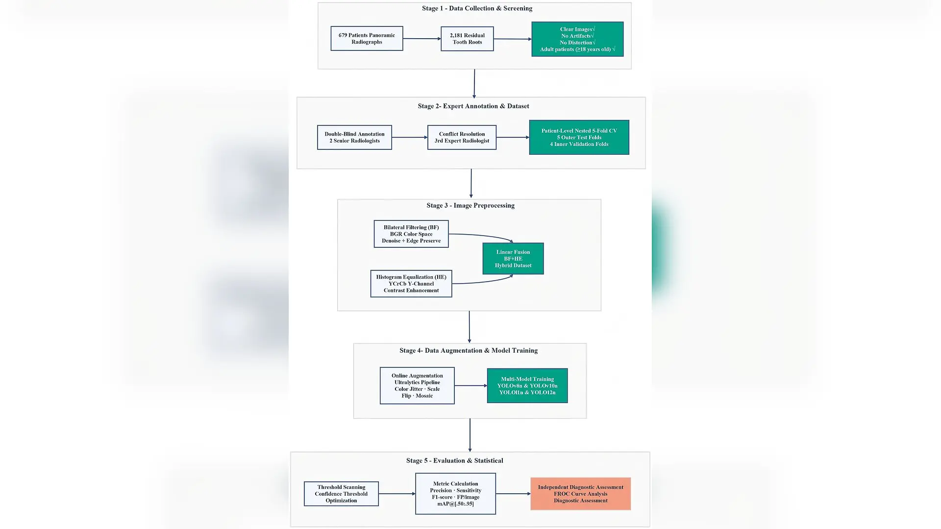
A tiny left-behind piece of a tooth root can trigger hidden bone infections or ruin a dental implant, but dentists frequently miss them on standard X-rays because the fragments blend seamlessly into the surrounding jawbone. To solve this diagnostic challenge, a team of researchers has evaluated lightweight artificial intelligence models to automatically spot these elusive dental remnants on panoramic dental X-rays.
Retained root fragments occur when a tiny piece of a tooth is left behind following a difficult extraction or when the top crown of a severely decayed tooth breaks off and the remaining roots stay trapped in the jawbone. If left alone, these fragments can form painful cysts and cause chronic inflammation that complicates future dental procedures. Traditional diagnosis relies heavily on human visual inspection of panoramic images, which is prone to human error because the tiny fragments often hide behind complex anatomical landmarks like the mandibular canal.
To build a reliable digital detection tool, the research team published in PLOS One a comprehensive evaluation of four nano-scale versions of the popular You Only Look Once computer vision framework. Specifically, the scientists tested YOLOv8n, YOLOv10n, YOLO11n, and YOLO12n to see which model performed best at pinpointing these hidden root fragments.
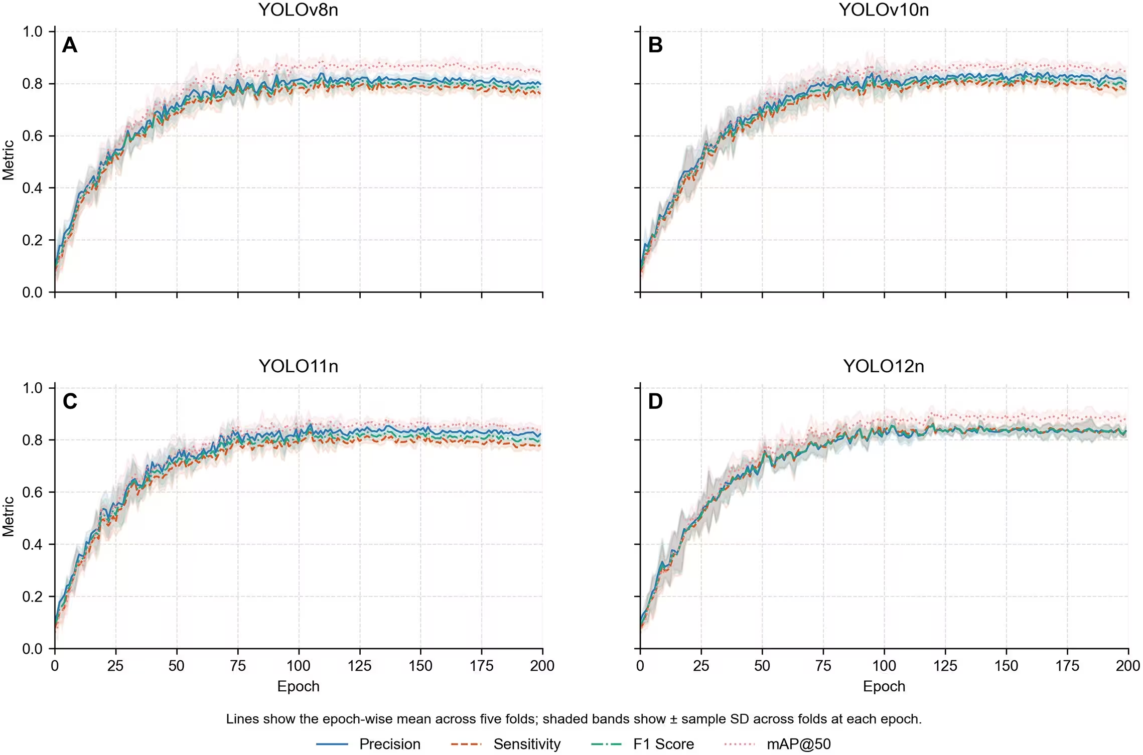
How the Artificial Intelligence Models Were Trained and Tested
The retrospective study utilized a curated dataset of 679 patient panoramic radiographs gathered between April 2021 and October 2025. Two experienced oral and maxillofacial radiologists manually annotated a total of 2,181 residual root fragments to establish a gold standard for training. To prevent data leakage and ensure trustworthy results, the authors implemented a strict patient-level nested five-fold cross-validation scheme.
Dental X-rays are notoriously low in contrast, making it difficult for computer algorithms to isolate faint edges against trabecular bone. To overcome this limitation, the researchers developed a hybrid image preprocessing pipeline that combined bilateral filtering to smooth out background noise while preserving sharp edges, along with histogram equalization on the luminance channel to enhance local contrast. The team fused these two approaches using weighted linear blending to ensure the images maintained a natural radiographic appearance.
Each of the four nano-scale models was trained for 200 epochs using identical computational settings. Because these are nano-scale architectures containing fewer than three million parameters, they are specifically designed to operate efficiently within resource-constrained environments like chairside computer systems or mobile dental units.
Which YOLO Model Found the Most Root Fragments?
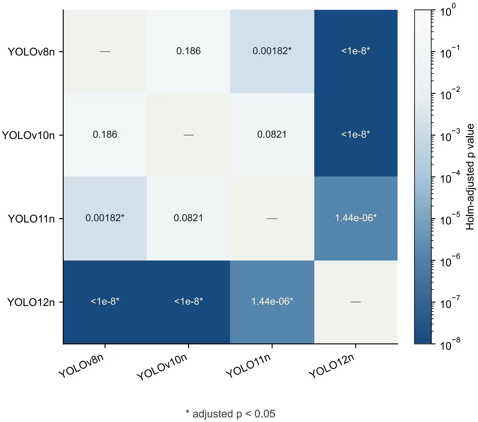
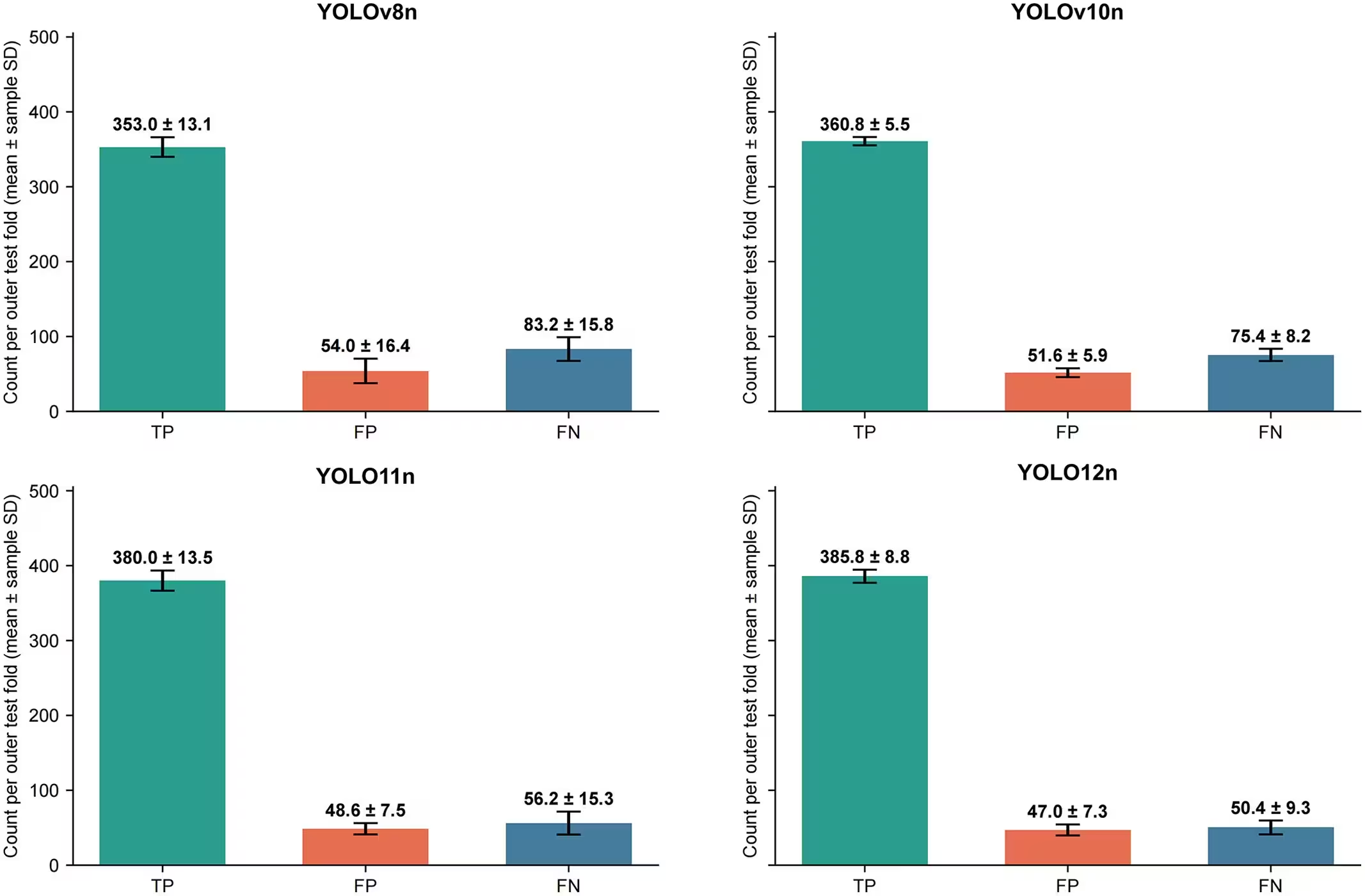
When evaluated across the held-out test folds, the newer attention-centric architectures significantly outperformed their predecessors. The newest model in the group, YOLO12n, achieved the highest overall detection performance, yielding a mean F1 score of 0.885 and a mean average precision at 0.5 threshold of 0.919. The F1 score is a statistical measure that balances both precision and sensitivity, where a score of 1.0 represents perfect accuracy.
YOLO11n followed closely behind as the second-best performing architecture, securing an F1 score of 0.874. Both top-performing models benefited from advanced spatial attention mechanisms, such as area attention and C2PSA modules, which helped the algorithms capture long-range spatial dependencies and distinguish thin root apices from surrounding bone structures. Meanwhile, the baseline YOLOv8n model recorded the lowest overall performance with an F1 score of 0.850.
Understanding the Limitations and Next Steps
Despite the high accuracy scores achieved in the laboratory, the authors noted several important limitations that must be addressed before clinical deployment. The study relied on a single-class design that only looked for root fragments, meaning it does not yet reflect the multi-class reality of busy dental clinics where patients present with multiple overlapping dental issues at once.
Furthermore, the dataset was constructed exclusively using X-rays that already contained at least one confirmed root fragment. Testing the software on healthy populations in routine screening settings could alter false-positive rates. The team emphasizes that extensive multi-center external validation and prospective clinical trials across unselected patient populations will be required before these lightweight tools can be safely integrated into everyday dental practice.
The research was published in PLOS One on October 23, 2026.
This article has been fact checked for accuracy, with information verified against reputable sources. Learn more about us and our editorial process.
Last reviewed on .
Article history
- Latest version
Reference(s)
- Ma, Chunyan., et al. “Comparative evaluation of YOLO models for retained root fragment detection.” PLOS One, vol. 21, no. 10, October 23, 2026, pp. e0358937 Public Library of Science (PLoS), doi: 10.1371/journal.pone.0358937. <https://doi.org/10.1371/journal.pone.0358937>.
Cite this page:
- Posted by Robert Brown