Human Stem Cell Embryo Models Reveal Surprising Structural Variability
Biotechnology

Human Stem Cell Embryo Models Reveal Surprising Structural Variability

Time-lapse imaging and single-colony sequencing expose hidden developmental differences in engineered human tissue models.
By Rohan Kumar
Published:
Email this Article
Human Stem Cell Embryo Models Reveal Surprising Structural Variability
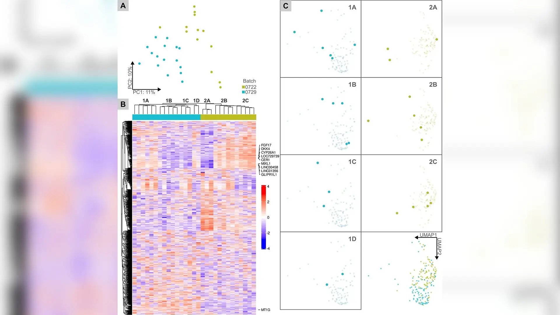
Transcriptomic profiles of single gastruloids are heterogeneous. (a) Principal component analysis (PCA) plot of gene expression profiles from single gastruloids. Proportions of variances indicated for PC1 and PC2. Hue corresponds to batch. (b) Heatmap displaying normalized relative expression of the top 1000 highly variable genes. Hierarchical clustering of both genes and samples (hue corresponds to batch) was performed using Pearson correlation distance with average linkage. Sample clusters and top 10 highly variable genes were labeled. (c) UMAP plots visualizing the dynamic latent representations of the sequenced gastruloids (large markers) in each subcluster and of the gastruloids in each batch (small markers). Hue corresponds to batch. For panels (a)–(c), N = 2 batches, n0722 = 13 gastruloids, and n0729 = 16 gastruloids were sequenced. Ian Jan et al. / APL bioengineering

Living human stem cell models reveal unexpected structural variability as they organize into early embryonic structures, raising important questions about the consistency of laboratory-grown tissues. Scientists at the University of Washington tracked thousands of developing human stem cell aggregates, known as gastruloids, and discovered profound differences in their physical patterning over a forty-eight hour window. The findings point to hidden sources of variability in human in vitro systems that could impact how researchers model early development and screen reproductive therapeutics.

Early human development involves intricate cellular coordination where a single misplaced signal can alter the fate of an entire tissue. Because studying natural human embryos carries severe technical and ethical barriers, researchers rely on human stem cell-based embryo models to replicate the earliest stages of formation. These two-dimensional structures self-organize into distinct germ layers that resemble the blueprint of an actual embryo, offering a scalable alternative to animal testing. However, the degree to which these engineered models vary from one another under identical laboratory conditions has remained largely unquantified until now.

How Tiny Magnetic Rafts Enabled Single-Cell Tracking

To capture how individual structures develop over time, the research team utilized an automated microraft platform consisting of hundreds of tiny, optically clear carriers embedded with magnetic edges. Human pluripotent stem cells expressing fluorescent reporters for SOX2, a key protein marking the early nervous system and pluripotency, were seeded onto these arrays and treated with bone morphogenetic protein 4 to trigger self-organization. The platform allowed the team to continuously image living structures every six hours for two days before mechanically isolating individual entities using a magnetic wand for downstream genetic analysis.
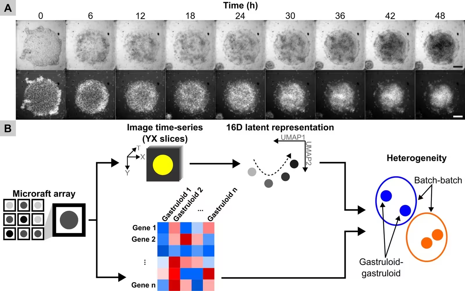
FIG. 1: Exploring single gastruloid heterogeneity. (a) Shown are sequential bright-field (upper row) and fluorescence (lower row) images using widefield microscopy of a single microraft possessing hPSCs expressing a SOX2-mCitrine reporter. BMP4 was added to the cultures at time 0 h to initiate gastruloid formation. Scale bars, 100 μm. (b) Schematic of the live time-series imaging and end point RNA-seq workflow. SOX2 dynamics and gene expression profiles of single gastruloids were acquired. 16D latent representations were formed from individual YX slices of imaging time-series (widefield microscopy) of each gastruloid and visualized via UMAP dimensionality reduction. Gastruloid heterogeneity across and within experimental batches was assessed using the image-based latent representations and sequence-based gene expression profiles.
Exploring single gastruloid heterogeneity. (a) Shown are sequential bright-field (upper row) and fluorescence (lower row) images using widefield microscopy of a single microraft possessing hPSCs expressing a SOX2-mCitrine reporter. BMP4 was added to the cultures at time 0 h to initiate gastruloid formation. Scale bars, 100 μm. (b) Schematic of the live time-series imaging and end point RNA-seq workflow. SOX2 dynamics and gene expression profiles of single gastruloids were acquired. 16D latent representations were formed from individual YX slices of imaging time-series (widefield microscopy) of each gastruloid and visualized via UMAP dimensionality reduction. Gastruloid heterogeneity across and within experimental batches was assessed using the image-based latent representations and sequence-based gene expression profiles. Credit: Ian Jan et al. / APL bioengineering

By applying self-supervised deep learning tools to thousands of time-lapse images, the researchers constructed low-dimensional mathematical representations that mapped the physical trajectory of each growing structure. The analysis, published in APL Bioengineering, revealed that while the stem cell colonies looked remarkably similar right after seeding, their structural trajectories diverged significantly as time progressed. While some structures formed neat, centralized rings of SOX2 activity, others developed irregular shapes or shifted away from expected symmetry.

What Genetic Sequencing Revealed About Hidden Batch Differences

To connect physical appearance with molecular activity, the team extracted RNA from twenty-nine individual structures across multiple experimental batches and performed bulk RNA sequencing. Principal component analysis and hierarchical clustering demonstrated that gene expression profiles separated cleanly by experimental batch, reflecting subtle batch-to-batch variations in cell culture dynamics. Among the most variable genes identified were critical developmental regulators such as CER1 and DKK4, which act as antagonists in major signaling pathways, alongside structural patterning genes like FGF17 and CYP26A1.

Interestingly, when the researchers attempted to directly correlate specific visual features from the imaging latent space with the expression levels of these top variable genes, no simple linear patterns emerged. This complexity suggests that single genes perform multiple overlapping functions during self-organization, meaning physical appearance and transcriptional output do not always map in a straightforward manner. The multimodal capability of the microraft technology successfully bridged the gap between live microscopy and molecular profiling, paving the way for more rigorous quality control in stem cell research.
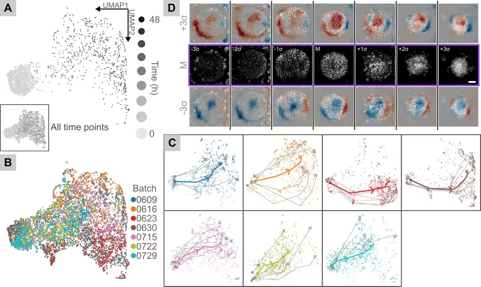
FIG. 2: Static latent representations learned from individual gastruloid images reveal heterogeneity. (a) UMAP plots visualizing 16D latent representations learned from individual static YX slices from imaging time-series of gastruloids. The main plot shows latent representations from initial and final time points (relative sizes of markers in the legend are not drawn to the same scale as markers in the plot), while the subplot shows latent representations from every time point. Shade within the gradient corresponds to the time point (6-h interval) during the 48-h differentiation protocol. (b) UMAP plot visualizing latent representations from every time point where hue corresponds to batch. (c) UMAP plots for each batch visualizing average temporal trajectories of all batch-specific latent representations and trajectories of five randomly sampled gastruloids. The start (circle) and end points (x) for each trajectory were labeled. Hue corresponds to batch. For panels (a)–(c), N = 7 batches. (d)
Static latent representations learned from individual gastruloid images reveal heterogeneity. (a) UMAP plots visualizing 16D latent representations learned from individual static YX slices from imaging time-series of gastruloids. The main plot shows latent representations from initial and final time points (relative sizes of markers in the legend are not drawn to the same scale as markers in the plot), while the subplot shows latent representations from every time point. Shade within the gradient corresponds to the time point (6-h interval) during the 48-h differentiation protocol. (b) UMAP plot visualizing latent representations from every time point where hue corresponds to batch. (c) UMAP plots for each batch visualizing average temporal trajectories of all batch-specific latent representations and trajectories of five randomly sampled gastruloids. The start (circle) and end points (x) for each trajectory were labeled. Hue corresponds to batch. For panels (a)–(c), N = 7 batches. (d) Credit: Ian Jan et al. / APL bioengineering

Why Developmental Variability Matters for Future Research

The discovery of deep structural and genetic heterogeneity within genetically identical stem cell lines carries significant implications for biomedical engineering. As regulatory agencies increasingly push to reduce reliance on animal testing, human in vitro models are taking center stage for drug screening and toxicity testing. However, if laboratory-grown models exhibit high intrinsic variability, researchers must carefully account for these differences in their experimental design to avoid misinterpreting drug responses or developmental defects.

The authors note that this study serves as a foundational proof-of-concept, limited by its sample size and the use of a single reporter cell line. Future investigations incorporating additional lineage markers and larger datasets will help determine whether this variability stems from intrinsic cellular noise or extrinsic microenvironmental factors. By embracing rather than ignoring this biological noise, scientists can gain a more realistic picture of the delicate mechanical forces shaping early human development.

The research was published in APL bioengineering.

Fact Checked

This article has been fact checked for accuracy, with information verified against reputable sources. Learn more about us and our editorial process.

Last reviewed on .

Article history

  • Latest version

Reference(s)

  1. Jan, Ian., et al. “Heterogeneous patterning behavior emerges from human stem cell-based embryo model.” APL Bioengineering, vol. 10, no. 4, November 12, 2026 AIP Publishing, doi: 10.1063/5.0329616. <https://doi.org/10.1063/5.0329616>.

Cite this page:

Kumar, Rohan. “Human Stem Cell Embryo Models Reveal Surprising Structural Variability.” BioScience. BioScience ISSN 2521-5760, 15 November 2026. <https://www.bioscience.com.pk/en/subject/biotechnology/heterogeneous-patterning-behavior-emerges-from-human-stem-cell-based-embryo-model>. Kumar, R. (2026, November 15). “Human Stem Cell Embryo Models Reveal Surprising Structural Variability.” BioScience. ISSN 2521-5760. Retrieved November 15, 2026 from https://www.bioscience.com.pk/en/subject/biotechnology/heterogeneous-patterning-behavior-emerges-from-human-stem-cell-based-embryo-model Kumar, Rohan. “Human Stem Cell Embryo Models Reveal Surprising Structural Variability.” BioScience. ISSN 2521-5760. https://www.bioscience.com.pk/en/subject/biotechnology/heterogeneous-patterning-behavior-emerges-from-human-stem-cell-based-embryo-model (accessed November 15, 2026).
End of the article