New Python Tool Reveals How Bacteria Build Complex 3D Biofilms
An open-source framework uncovers how genetics and nutrients jointly shape the hidden multi-scale architecture of microbial communities.
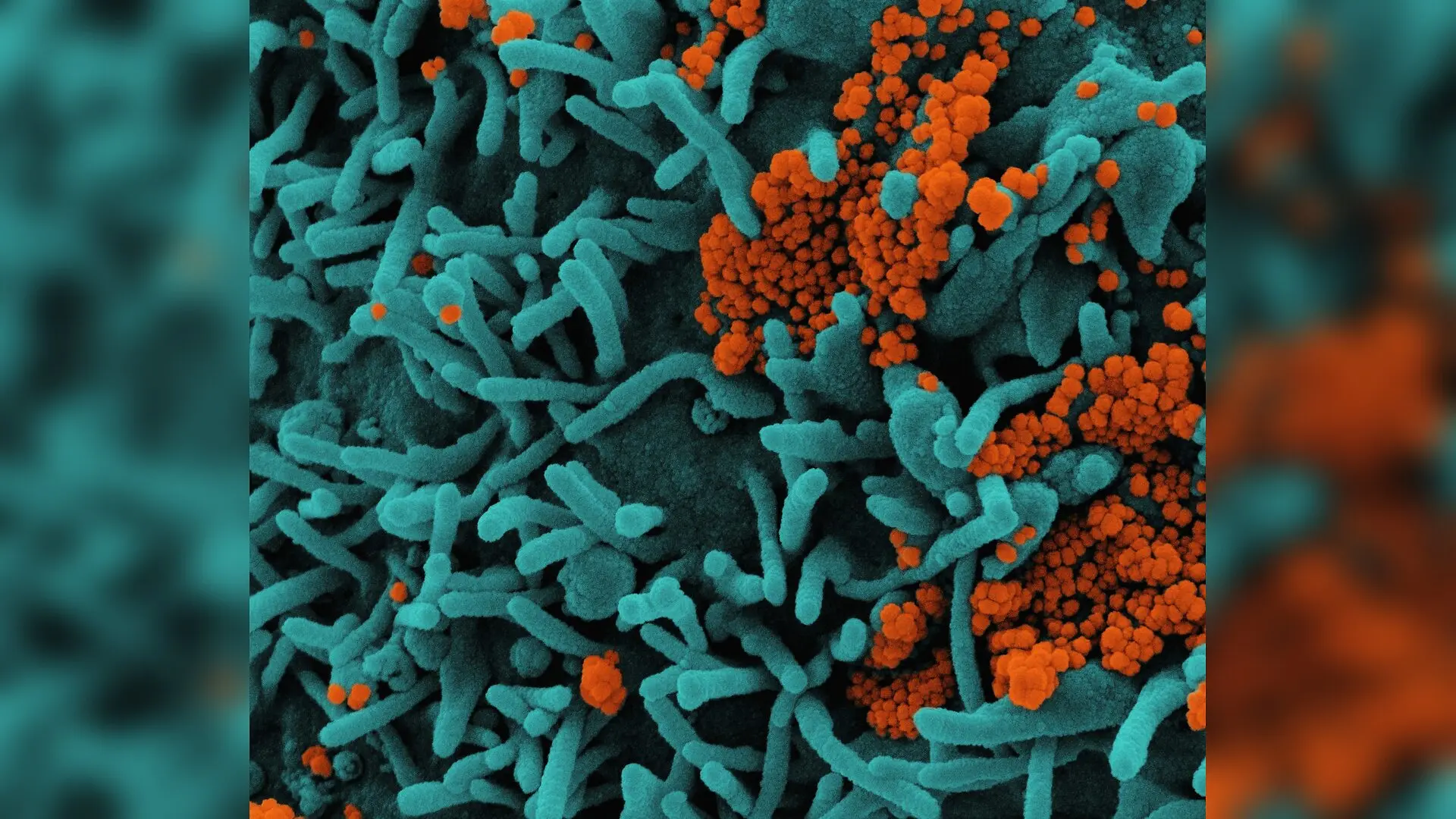
Microbial communities often form complex three-dimensional structures known as biofilms, which protect cells from environmental stress and antimicrobial treatments. Understanding how these communities build their physical shape has been difficult because traditional software tools rely on simple measurements like total volume or two-dimensional projections that miss crucial internal details. To solve this problem, a new open-source Python tool called Analysis of Biofilm Complexity in 3D (ABC3D) has been developed to automatically extract volumetric fractal, textural, and statistical descriptors from confocal microscopy images.
How the Open-Source Python Tool Quantifies Biofilm Architecture
The research is published in Royal Society Open Science, detailing an automated pipeline that processes volumetric image data to map structural heterogeneity. Confocal laser scanning microscopy captures three-dimensional stacks of bacterial macrocolonies, which are then segmented to isolate the biomass from the background. Instead of relying on a single bulk measurement, the software computes multiple parameters simultaneously, including box-counting fractal dimension, lacunarity to measure porosity, grey-level co-occurrence matrix texture features, Shannon entropy, and relative wavelet sub-band energies.
To test the framework, the author repurposed previously published imaging datasets of Escherichia coli macrocolonies. These datasets included the parental strain BW25113 alongside three deletion mutants, namely delta-amiA, delta-ompR, and delta-ydgD, grown on nutrient-rich lysogeny broth agar for 24 hours or minimal M9 agar for 48 hours. Principal component analysis of the resulting feature matrices showed that the first two principal components captured 75.5 percent of the total structural variance across all tested conditions.
Nutrient Conditions and Genetics Shape Coordinated Microstructure
Statistical evaluations revealed that both the growth medium and the genetic background significantly influence biofilm architecture, with a strong interaction effect. For instance, the delta-ydgD mutant exhibited large shifts in principal component scores between the rich and minimal media, whereas other strains showed different trajectories. This non-uniform response demonstrates that architectural reorganization is a coordinated multi-scale phenotype rather than a simple scaling of overall size.
The analysis also confirmed that nutrient changes alter structural descriptors across multiple complementary families. Rather than experiencing a uniform increase in complexity, specific mutants redistributed their textural and spatial-frequency features depending on the available nutrients. This multi-scale approach provides a clearer picture of how bacteria adapt their physical microenvironment in response to environmental pressures.
Limitations and Future Prospects for Structural Phenotyping
Despite its utility, the framework has certain technical boundaries that stem from the nature of light microscopy. Confocal imaging typically provides lower axial resolution than lateral resolution, and thick macrocolony specimens can suffer from light scattering and depth-dependent signal attenuation. Furthermore, the global thresholding method used for segmentation may occasionally miss subtle structural variations in dense layers.
The author notes that future updates could incorporate multi-level spectral analysis and fully three-dimensional texture calculations to refine the measurements further. By bridging the gap between genetics, environment, and physical shape, this software offers researchers a standardized way to evaluate how structural organization impacts microbial function and stress resistance.
The research was published in Royal Society open science on November 11, 2026.
This article has been fact checked for accuracy, with information verified against reputable sources. Learn more about us and our editorial process.
Last reviewed on .
Article history
- Latest version
Reference(s)
- McConnell, Gail. “Analysis of biofilm complexity in 3D: an open-source framework for quantitative fractal, textural and statistical analysis of colony biofilm morphology in three dimensions.” Royal Society Open Science, vol. 13, no. 11, November 11, 2026 The Royal Society, doi: 10.1098/rsos.260853. <https://doi.org/10.1098/rsos.260853>.
Cite this page:
- Posted by Robert Brown