Surgery and Anesthesia Trigger Hidden Brain Fat Disruption in APOE4 Carriers
Biology

Surgery and Anesthesia Trigger Hidden Brain Fat Disruption in APOE4 Carriers

Mouse study reveals how a common volatile anesthetic and surgical stress induce glial lipid breakdown, electrical circuit dysfunction, and delayed cognitive decline.
By Priya Sharma
Published:
Email this Article
Surgery and Anesthesia Trigger Hidden Brain Fat Disruption in APOE4 Carriers
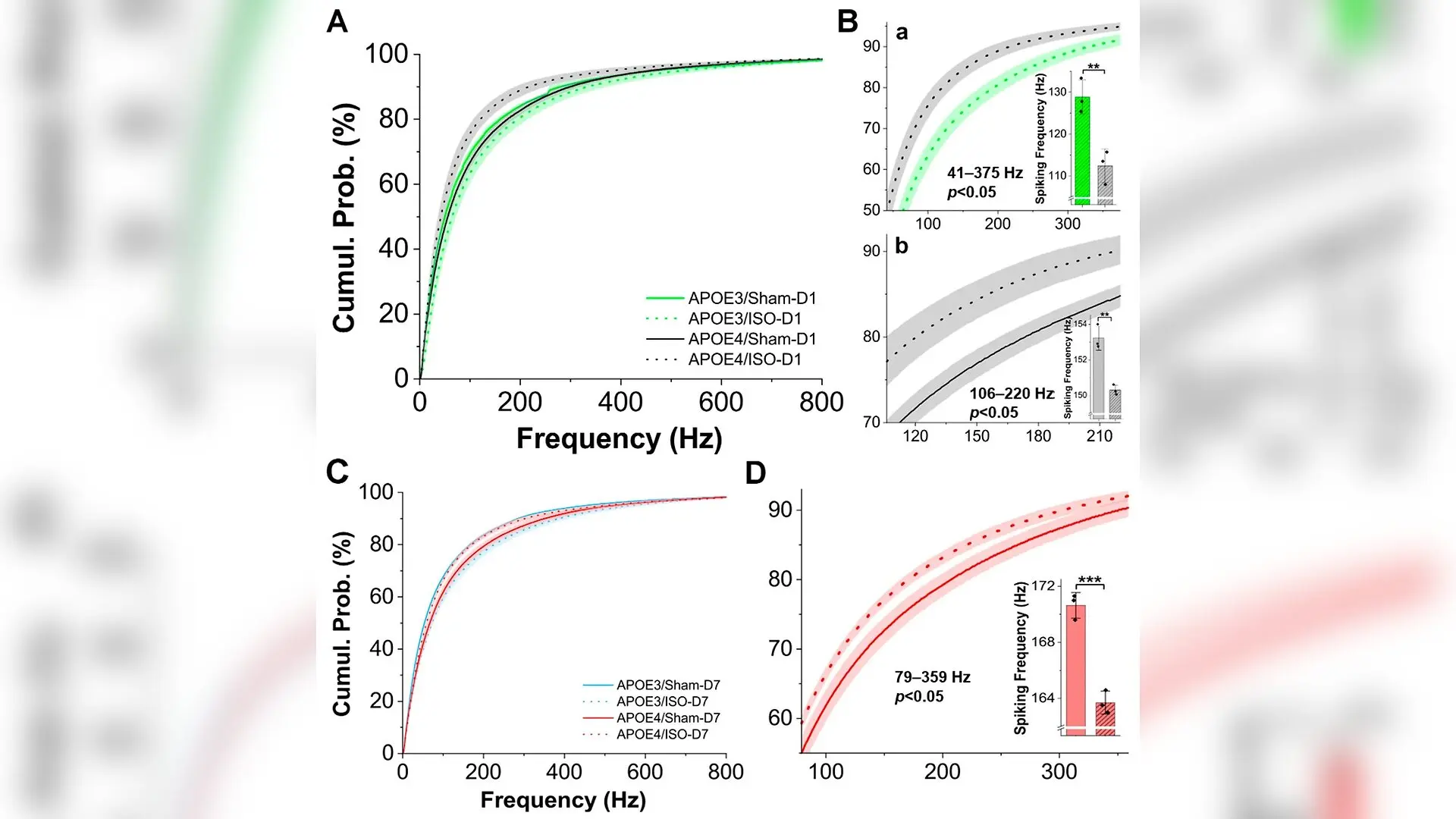
Isoflurane (ISO) effects on neuronal excitability in the OB mitral cell layer on post-ISO day 1 (D1) and 7 (D7). Line graphs were compared using the number of animals as the sample size and with the false discovery rate (FDR) correction to identify the frequency ranges with statistical significance. Bar graphs represent the averaged spike frequency within the corresponding frequency range with statistical significance from line graphs on the left and were analyzed using a two-sample t-test, with the animal as the experimental unit. (A) Comparison of the cumulative probability of spiking frequency on D1 post-ISO exposure among four groups of mice: (1) APOE3/sham (APOE3/Sham-D1, solid green curve, n = 3 mice with 7 recordings), (2) APOE3/ISO (APOE3/ISO-D1, dashed green curve, n = 3 mice with 6 recordings), (3) APOE4/sham (APOE4/Sham-D1, solid black curve, n = 3 mice with 6 recordings), (4) APOE4/ISO (APOE4/ISO-D1, dashed black curve, n = 3 mice with 6 recordings). (B) Enlarged view of A Yun Li et al. / Cells

A common genetic variant carried by roughly 25 to 30 percent of adults makes the brain uniquely vulnerable to routine surgery and general anesthesia, according to new research in mice. When young adult male mice carrying the Alzheimer’s disease genetic risk factor apolipoprotein E4, or APOE4, underwent a standard two-hour surgical and anesthesia procedure, they experienced profound disruptions in brain cell fat regulation, followed by lasting sensory and cognitive declines.

The study, published in Cells, sheds light on why some individuals suffer from persistent mental fog or memory difficulties after undergoing operations that use common volatile anesthetics like isoflurane. While millions of people undergo surgery safely every year, researchers have long sought to understand why certain patients develop postoperative neurocognitive dysfunction or face an elevated long-term risk of neurodegenerative diseases.

How Brain Fat Chemistry Derails After Surgery

To investigate what happens inside the brain during the perioperative period, which encompasses the time before, during, and after surgery, the research team focused on glial cells. These specialized support cells, which include astrocytes and microglia, maintain chemical balance and manage waste clearance in the brain. They are also heavily responsible for processing lipids, which are essential fats that make up more than half of the brain’s dry weight. Using humanized knock-in mice engineered to carry either the protective APOE3 gene or the high-risk APOE4 gene, the team examined what happened seven days after the animals underwent a two-hour isoflurane exposure paired with a exploratory laparotomy surgery to simulate surgical stress.
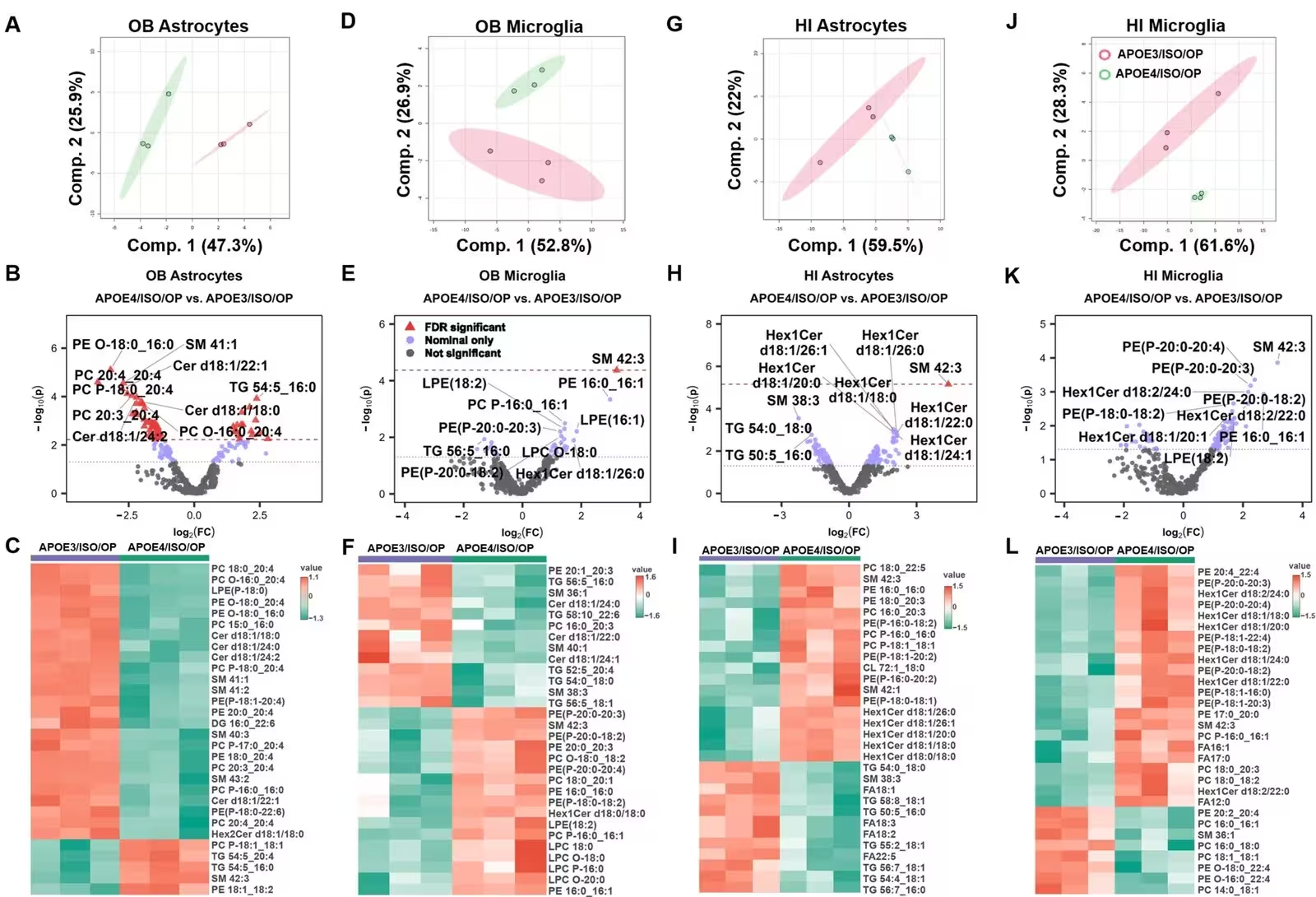
Figure 1: APOE4 drives heightened lipid dysregulation in olfactory bulb (OB) and hippocampal (HI) astrocytes and microglia 7 days after isoflurane (ISO) exposure and laparotomy operation (OP). (A–C) OB astrocytes, (D–F) OB microglia, (G–I) HI astrocytes, and (J–L) HI microglia from APOE4/ISO/OP vs. APOE3/ISO/OP mice at 7 days post-exposure. In each set, Partial least squares discriminant analysis (PLS-DA) illustrates sample clustering. Volcano plots display log2 fold changes and nominal p-values, with red triangles indicating lipids meeting the Benjamini–Hochberg false discovery rate (FDR)-adjusted significance threshold (adjusted p ≤ 0.05), purple points indicating nominal significance only (p < 0.05), and grey points indicating non-significant lipids. The red dotted horizontal line represents the nominal p-value cutoff corresponding to the FDR-adjusted significance threshold for each comparison. Heatmaps show the top 30 lipids selected by nominal Student’s t-test p-values for exploratory visua
APOE4 drives heightened lipid dysregulation in olfactory bulb (OB) and hippocampal (HI) astrocytes and microglia 7 days after isoflurane (ISO) exposure and laparotomy operation (OP). (A–C) OB astrocytes, (D–F) OB microglia, (G–I) HI astrocytes, and (J–L) HI microglia from APOE4/ISO/OP vs. APOE3/ISO/OP mice at 7 days post-exposure. In each set, Partial least squares discriminant analysis (PLS-DA) illustrates sample clustering. Volcano plots display log2 fold changes and nominal p-values, with red triangles indicating lipids meeting the Benjamini–Hochberg false discovery rate (FDR)-adjusted significance threshold (adjusted p ≤ 0.05), purple points indicating nominal significance only (p < 0.05), and grey points indicating non-significant lipids. The red dotted horizontal line represents the nominal p-value cutoff corresponding to the FDR-adjusted significance threshold for each comparison. Heatmaps show the top 30 lipids selected by nominal Student’s t-test p-values for exploratory visua Credit: Yun Li et al. / Cells

The lipidomic profiling revealed striking differences. Microglia and astrocytes from the olfactory bulbs and hippocampi of APOE4 mice showed severe, widespread lipid dysregulation compared to their APOE3 counterparts. The researchers observed abnormal remodeling across multiple fat classes, including triglycerides, phospholipids, and sphingomyelins. In healthy brains, support cells store excess fats safely in lipid droplets to prevent toxicity. The depletion of specific triglycerides in the APOE4 mice suggests that this vital buffering system fails under surgical stress, leaving brain cells vulnerable to metabolic strain.

Electrical Storms and Circuit Breakdowns in the Olfactory Bulb

These molecular disruptions did not stay confined to chemistry. Electrophysiological recordings in awake, head-fixed animals revealed that isoflurane exposure immediately altered electrical firing patterns in the olfactory bulb, a brain region responsible for processing smells and one of the earliest areas affected in Alzheimer’s disease. On day one following the procedure, mitral output neurons in APOE4 mice exhibited suppressed firing frequencies and disrupted network oscillations in specific frequency bands, contrasting sharply with the hyperexcitability seen in APOE3 control animals.

Patch-clamp recordings conducted on brain slices seven days later confirmed that synaptic transmission between neurons was fundamentally altered. In the olfactory bulb external tufted cells of APOE4 mice, baseline excitatory signaling was elevated while inhibitory control was weakened, skewing the delicate balance required for normal neural communication. In contrast, hippocampal neurons showed different patterns of disruption, proving that anesthetics and surgical stress trigger distinct, region-specific vulnerabilities depending on a patient’s genetic makeup.
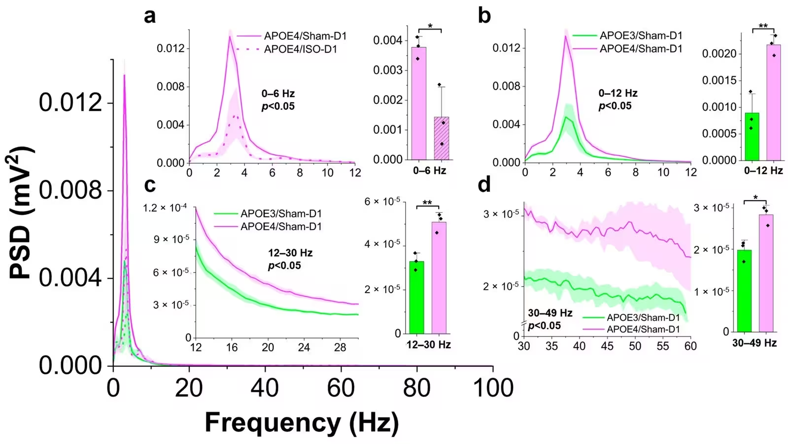
Figure 3: Isoflurane (ISO) effects on local field potential (LFP) oscillations of on post-treatment days 1 (D1) and 7 (D7). Line graphs were compared using the number of animals as the sample size, with the false discovery rate (FDR) correction applied, whereas the bar graphs represent the averaged power spectral density (PSD) within the corresponding frequency range with statistical significance on the left and were analyzed using a two-sample t-test with the animal as the experimental unit. The main panel shows the comparison of the PSD of LFP oscillatory activities on D1 post- ISO-exposure among four groups of mice (n-3 mice/group): (1) APOE3/Sham (APOE3/Sham-D1, solid green curve, 7 recordings), (2) APOE3/ISO (APOE3/ISO-D1, dashed green curve, 6 recordings), (3) APOE4/Sham (APOE4/Sham-D1, solid purple curve, 6 recordings), and (4) APOE4/ISO (APOE4/ISO-D1, dashed purple curve, 6 recordings). Inset (a) shows the comparison between APOE4 ISO and APOE4 sham groups on post- ISO-exposure day 1 (D1
Isoflurane (ISO) effects on local field potential (LFP) oscillations of on post-treatment days 1 (D1) and 7 (D7). Line graphs were compared using the number of animals as the sample size, with the false discovery rate (FDR) correction applied, whereas the bar graphs represent the averaged power spectral density (PSD) within the corresponding frequency range with statistical significance on the left and were analyzed using a two-sample t-test with the animal as the experimental unit. The main panel shows the comparison of the PSD of LFP oscillatory activities on D1 post- ISO-exposure among four groups of mice (n-3 mice/group): (1) APOE3/Sham (APOE3/Sham-D1, solid green curve, 7 recordings), (2) APOE3/ISO (APOE3/ISO-D1, dashed green curve, 6 recordings), (3) APOE4/Sham (APOE4/Sham-D1, solid purple curve, 6 recordings), and (4) APOE4/ISO (APOE4/ISO-D1, dashed purple curve, 6 recordings). Inset (a) shows the comparison between APOE4 ISO and APOE4 sham groups on post- ISO-exposure day 1 (D1 Credit: Yun Li et al. / Cells

From Early Smell Loss to Delayed Memory Decline

Behavioral tracking over a twelve-week period revealed a distinct chronological timeline of neurological impairment. APOE4 mice subjected to anesthesia and surgery developed persistent olfactory memory deficits starting around day 11, struggling to recognize previously encountered scents. They also exhibited temporary difficulties in locating buried food, signaling a temporary loss of olfactory sensitivity. Early motor coordination deficits resolved within the first two weeks, but a more concerning delayed symptom emerged: by day 86, the APOE4 mice displayed significant spatial working-memory impairments in Y-maze testing.

Long-term transcriptomic profiling of the olfactory bulb at 90 days post-exposure identified lasting alterations in genes related to calcium ion transport and cellular metabolism. Although the overall number of differentially expressed genes was relatively modest compared to baseline genetic differences, the persistence of these molecular shifts highlights how a single early surgical exposure can leave a permanent imprint on genetically susceptible brain tissue. The authors note that while these findings provide valuable mechanistic insights, further research involving female cohorts and additional anesthetic agents will be necessary to fully map out human clinical risks.

The research was published in Cells on November 9, 2026.

Fact Checked

This article has been fact checked for accuracy, with information verified against reputable sources. Learn more about us and our editorial process.

Last reviewed on .

Article history

  • Latest version

Reference(s)

  1. Li, Yun., et al. “Isoflurane and Surgery Aggravate APOE4-Dependent Lipid Dysregulation, Neural Dysfunction, and Neurological Impairment in Male Mice.” Cells, vol. 15, no. 21, November 9, 2026, pp. 2038 MDPI AG, doi: 10.3390/cells15212038. <https://doi.org/10.3390/cells15212038>.

Cite this page:

Sharma, Priya. “Surgery and Anesthesia Trigger Hidden Brain Fat Disruption in APOE4 Carriers.” BioScience. BioScience ISSN 2521-5760, 16 November 2026. <https://www.bioscience.com.pk/en/subject/biology/isoflurane-and-surgery-aggravate-apoe4-dependent-lipid-dysregulation-neural-dysfunction-and-neurological-impairment-in-male-mice>. Sharma, P. (2026, November 16). “Surgery and Anesthesia Trigger Hidden Brain Fat Disruption in APOE4 Carriers.” BioScience. ISSN 2521-5760. Retrieved November 16, 2026 from https://www.bioscience.com.pk/en/subject/biology/isoflurane-and-surgery-aggravate-apoe4-dependent-lipid-dysregulation-neural-dysfunction-and-neurological-impairment-in-male-mice Sharma, Priya. “Surgery and Anesthesia Trigger Hidden Brain Fat Disruption in APOE4 Carriers.” BioScience. ISSN 2521-5760. https://www.bioscience.com.pk/en/subject/biology/isoflurane-and-surgery-aggravate-apoe4-dependent-lipid-dysregulation-neural-dysfunction-and-neurological-impairment-in-male-mice (accessed November 16, 2026).
End of the article